From Light to Sight: Human Color Perception
Curious about how the human eye perceives and distinguishes colors? Human color perception plays a crucial role in this ability, letting us see all the beautiful shades in the world around us, turning light into colorful images that our brains understand.
The human eye is one of nature’s most amazing creations. Each day, this incredible organ processes millions of color signals, transforming simple wavelengths of light into the rich, detailed world we experience. Scientists estimate that the human eye can distinguish approximately 10 million different colors, creating a visual experience that has fascinated researchers for centuries.

The human eye detects color much like a digital camera. Light bounces off objects, enters your eye, and triggers special cells that sense red, green, or blue. Your brain interprets these signals. About 4-5% of people experience color vision deficiency, as shown below:
| Population Group | Prevalence of Color Vision Deficiency |
|---|---|
| Males | 5-8% |
| Females | 0.4-1% |
| Overall | 4-5% |
This process is called human eye color detection and differentiation.
Human Eye Color Detection and Differentiation
What is Color?
When you look at a leaf, a flower, or even the sky, you see a world full of colors. But what is color? In science, color is not just something objects possess. Instead, color is a perception that your brain creates when light interacts with your eyes. Light travels in waves, and each wave has a different length. These wavelengths fall within the visible spectrum, which spans approximately 400 to 700 nanometers. When sunlight hits an object, some wavelengths are absorbed, and others are reflected. The human eye color detection and differentiation process begins when light reflects off objects and enters your eyes. The reflected light triggers special cells called cones and rods. Your brain then interprets these signals as colors.
Isaac Newton demonstrated that sunlight is made up of all the visible colors. He used a prism to split sunlight into a rainbow, proving that white light is a mix of many colors. Goethe later pointed out that color is also a personal experience. You and your friend might see the same light, but your brains could interpret the colors a bit differently. This makes the nature of color both a physical and a personal thing.
A Journey through the Human Eye’s Structure
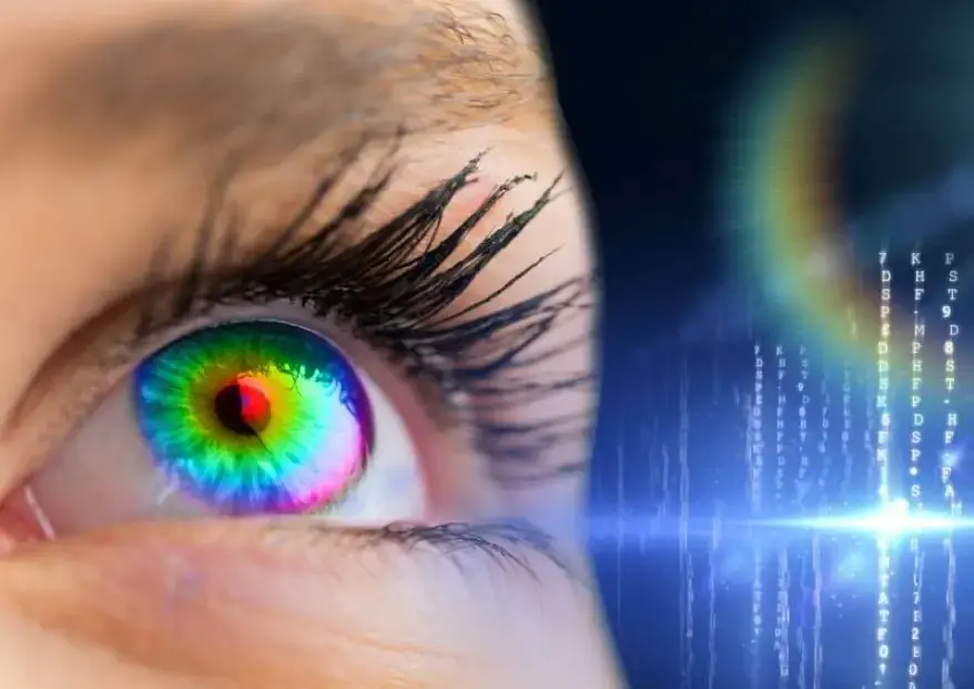
The journey of light into your eye is a key part of color detection and differentiation. First, light passes through the cornea, the clear front surface of your eye. The cornea bends the light and helps focus it. Next, the light moves through the pupil, which is the black opening in the center of your eye. The iris, the colored part around the pupil, controls how much light enters by making the pupil bigger or smaller. After that, the light goes through the lens. The lens changes shape to focus the light even more, making sure it lands on the right spot at the back of your eye. The light then travels through a jelly-like substance called the vitreous humor before reaching the retina. The retina is where the magic of color detection and differentiation truly happens.
| Structure | Role in color detection and differentiation |
|---|---|
| Cornea | Bends and focuses incoming light onto the retina. |
| Pupil/Iris | Regulates the amount of light entering the eye by adjusting pupil size. |
| Lens | Fine-tunes focus by changing shape to direct light onto the retina. |
| Vitreous Humor | Transparent gel that maintains eye shape and allows light to pass through to the retina. |
| Retina | Converts focused light into electrical signals for the brain to process visual information. |

Rods and Cones: The Two Classes of Photoreceptors
Once light reaches your retina, two types of cells get to work: rods and cones. Each cell has its own different role. Rods help you see in dim light and are very sensitive, but they do not detect color. They are mostly found in the outer parts of your retina. Cones, on the other hand, are responsible for color vision and sharp detail. They work best in bright light and are packed tightly in the center of your retina, called the fovea. The process of human eye color detection and differentiation relies on both rods and cones, but only cones let you see the full range of colors.
| Aspect | Rods | Cones |
|---|---|---|
| Sensitivity | High; work in dim light | Low; work in bright light |
| Vision Type | Black-and-white | Color (red, green, blue) |
| Location | Peripheral retina | Central retina (fovea) |
| Function | Night and peripheral vision | Daytime and color vision |
| Visual Acuity | Low | High |
| Response Time | Slow | Fast |
Trichromatic Vision: Seeing a Rainbow
In 1802, Thomas Young wrote: “If we seek for the simplest arrangement… we may suppose three distinct sensations only to be excited by the rays of the three principal pure colors—the red, the green, and the violet.” This profound insight later became the Trichromatic Theory of Vision.
The Three Types of Cones
Humans have three types of cones in the retina. Each type responds best to a different part of the visible spectrum:
- Firstly, S-cones (short-wavelength): Peak sensitivity around 420–445 nm, respond to blue light.
- Secondly, M-cones (medium-wavelength): Peak sensitivity around 530–543 nm, respond to green light.
- L-cones (long-wavelength): Peak sensitivity around 560–566 nm, respond to green light.
These cones work together in a process called trichromatic vision. When light hits your retina, each cone type sends a signal to your brain based on how much it is activated. Your brain compares these signals to figure out the exact color. For example, if both your L-cones and M-cones are highly active, you might see yellow. This teamwork between cones is a key part of human color perception, enabling us to distinguish subtle color variations. If you lose one type of cone, you may experience color blindness, which shows how important each cone is for color detection and differentiation.
A World Full of (and Beyond )Color
Expanding Human Color Perception
Visible Spectrum and Invisible Colors
The limits of human color perception are defined by our biology, preventing us from seeing beyond the visible spectrum. The visible spectrum for humans ranges from about 380 to 700 nanometers.
| Color | Wavelength Range (nm) |
|---|---|
| Violet | 380 – 450 |
| Deep Blue | 450 – 484 |
| Light Blue | 484 – 500 |
| Green | 500 – 565 |
| Yellow | 565 – 590 |
| Orange | 590 – 625 |
| Red | 625 – 700 |
Light with wavelengths shorter than violet is called ultraviolet. Light with wavelengths longer than red is called infrared. Human eyes cannot see these colors because photopigments do not respond to them. Moreover, the process of detecting and differentiating human eye colors is limited to the visible spectrum. Special technology can sometimes help you “see” ultraviolet or infrared, but your eyes alone cannot detect them.
How Many Colors Can We See?
You might wonder how many colors your eyes can tell apart. Most people can see about one million distinct colors. The incredible range of shades we can detect is a result of the complexity of human color perception. This is possible because each of your three cone types can detect around 100 different shades. When you combine all the possible combinations, you get a huge number of colors. Some rare people, usually women, have a fourth type of cone and can see up to 100 million colors. However, this is not common. The number of colors you can see depends on the structure of your eye, the lighting, and even how your brain processes signals. The process of human eye color detection and differentiation is truly amazing, letting you experience a colorful world every day.
Human Color Perception and Differences
Brain Processing of Color Signals
Your brain plays a crucial role in human color perception. When light hits the cones in your retina, these cells turn the light into electrical signals. Special molecules, such as ELFN2, help cones connect with cone-specific neurons. This connection ensures that each signal travels along the correct pathway. Your brain receives these signals and compares the activity from the three types of cones. To begin with, the primary visual cortex (V1) starts the process by registering the presence and intensity of different wavelengths. After that, areas like V2, V3, and V4 handle more complex color processing and help you recognize color in different lighting. Furthermore, the inferior temporal cortex stores your color knowledge and helps you remember colors. Altogether, these brain regions create your experience of color.

Why People See Colors Differently
Individual Variations in Human Color Perception
You may notice that sometimes people disagree about the color of an object. Genetic differences play a big part in this. Small changes in the genes that control cone photopigments can shift the sensitivity of your cones. Therefore, this means you might see a shade of green slightly differently than someone else. The number and arrangement of cones also vary. Some people have more L-cones (red-sensitive), while others have more M-cones (green-sensitive). The table below shows how these differences affect color vision:
| Aspect | Description |
|---|---|
| Cone Types in Normal Vision | Three types (L, M, S) allow full color vision. |
| Dichromats | Missing one cone type, see fewer colors. |
| Anomalous Trichromats | Have cones with shifted sensitivity, see colors differently. |
| Cone Proportions | S-cones are stable (~7.5%), L/M ratio varies widely among individuals. |
Iris color does not affect your ability to detect colors. Studies show that people with different iris colors perform equally well on color detection tasks.
Color Vision Anomalies
Color vision anomalies are common, especially among males. Most cases involve trouble seeing red and green shades.
| Color Vision Anomaly | Prevalence Rate | Notes |
|---|---|---|
| Red-Green Color Blindness | 98% of all cases | Most common globally |
| Deuteranomaly | 2.32% of the population | Most commonly affects the green cone |
| Protanomaly | 0.54% of the population | Affects the red cone |
| Tritanomaly | 0.02% of the population | Rare, affects blue cone |
| Achromatopsia | ~1 in 30,000 people | Complete color blindness, very rare |
Color vision anomalies can affect your daily life, especially in jobs that require color matching or reading color-coded information. Many people with color vision deficiency find ways to adapt, but early screening and support can help improve quality of life.

Conclusion
You experience color because your eye and brain work together in a series of steps:
- Light reflects off objects and enters your eye.
- Specialized cells in your retina—rods and cones—detect this light.
- Cones send signals to your brain, which interprets them as color.
Thus, you get to know how complex and unique your vision is. You see millions of colors, thanks to the teamwork of your eye and brain, which is a remarkable demonstration of human color perception. The invisibility of certain colors to human eyes is a direct consequence of biological and physical boundaries. Our eyes are equipped to detect only a specific segment of the electromagnetic spectrum. Appreciate this amazing ability every day!
References
- Bramão, I., Faísca, L., Forkstam, C., Reis, A., & Petersson, K. M. (2010). Cortical Brain Regions Associated with Color Processing: An FMRi Study. The Open Neuroimaging Journal, 4, 164–173. https://doi.org/10.2174/1874440001004010164
- Kawamura, S., & Tachibanaki, S. (2012). Explaining the functional differences of rods versus cones. Wiley Interdisciplinary Reviews Membrane Transport and Signalling, 1(5), 675–683. https://doi.org/10.1002/wmts.8
- Emery, K. J., & Webster, M. A. (2019). Individual differences and their implications for color perception. Current Opinion in Behavioral Sciences, 30, 28–33. https://doi.org/10.1016/j.cobeha.2019.05.002
- Zhang, F., Kurokawa, K., Bernucci, M. T., Jung, H. W., Lassoued, A., Crowell, J. A., Neitz, J., Neitz, M., & Miller, D. T. (2021). Revealing how color vision phenotype and genotype manifest in individual cone cells. Investigative Ophthalmology & Visual Science, 62(2), 8. https://doi.org/10.1167/iovs.62.2.8
Additionally, to stay updated with the latest developments in STEM research, visit ENTECH Online. Basically, this is our digital magazine for science, technology, engineering, and mathematics. Furthermore, at ENTECH Online, you’ll find a wealth of information.


