Tissue engineering (TE) is the science of building replacement body parts in a lab.
Scientists Grew a Working Esophagus in a Lab from a patient’s own cells
Tissue Engineering Careers
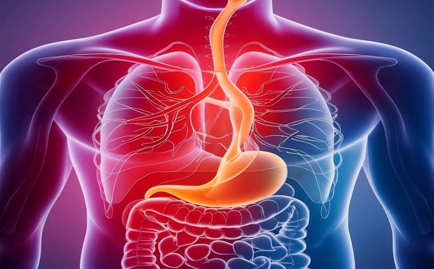
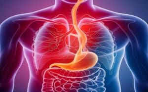
Imagine being born without a fully formed food pipe. You cannot swallow. You cannot eat normally. All of a sudden, your entire life revolves around surgeries and feeding tubes. This is the reality for many babies born with esophageal atresia (EA) — a condition where the esophagus does not connect properly. At first, doctors try to stitch the gaps. But in about 10% of cases, the gap is simply too large. At this point, at least so far, there were very few good options.
What Is Tissue Engineering, and Why Does It Matter?
- Tissue engineering (TE) is the science of building replacement body parts in a lab. It combines biology, materials science, and engineering. To enumerate the basic steps: scientists take a scaffold (a structural frame), seed it with living cells, grow it in a special machine called a bioreactor, and then implant it into the body.
- The esophagus needs muscles that actively push food down through wave-like movements called peristalsis. Growing that kind of muscle is incredibly difficult.
How Did They Build It?
The researchers used a decellularized porcine (pig) scaffold. To say it differently, they took a pig’s esophagus and stripped away all the pig cells. What remained was a clean, protein-rich framework — essentially an empty biological skeleton.
After that, they injected two types of the animal’s own cells into this scaffold:
- Mesoangioblasts (MABs) — pericyte-like cells with the ability to become muscle
- Fibroblasts (FBs) — cells that support tissue structure and matrix remodeling
They used a 7:3 ratio of MABs to fibroblasts. What’s more, they microinjected these cells at 120 precise points along the scaffold. So as to ensure the cells matured properly before surgery, they placed the entire graft inside a bioreactor — a chamber that mimics body conditions — for one week.
What the Bioreactor Did
The bioreactor changed the cells in a remarkable way. Seeing that the cells faced a low-oxygen environment inside the bioreactor, they switched on proangiogenic genes — genes that help attract new blood vessels. In similar fashion to how a seed prepares itself for harsh soil before planting, the cells primed themselves to survive and grow once inside a living body.
What Happened After Implantation?
The team surgically replaced a 2.5-centimetre section of the esophagus in eight minipigs, modeling the size of a pediatric patient. All animals received water immediately after surgery and began eating the next day.
Safety Results
All eight animals survived the first 30 days. At this point, that alone was a significant achievement. While this may be true that some complications arose — including polyp formation and occasional narrowing — these were manageable through standard endoscopic procedures. What’s more, these complications closely matched what doctors already see in real clinical practice for children with this condition.
Long-Term Outcomes
Five of eight animals (63%) reached the planned six-month endpoint. These five animals were eating normally. They showed normal weight gain compared to healthy reference minipigs. In due time, and with increasing complexity, the grafts developed organized tissue layers including epithelium, submucosa, and muscularis — layers that look just like a normal esophagus.
The Peristalsis Breakthrough
At last, and most critically: the grafts showed secondary peristalsis. Scientists used a special probe called high-resolution impedance manometry (HRIM) to measure pressure waves. In five of seven tested animals, the grafts produced real, coordinated muscle contractions. In essence, the lab-grown food pipe was actually squeezing and pushing food, just like a normal esophagus does.
Why This Is a Big Deal for Medicine
All in all, this study achieves several “firsts” at once. To sum up the key breakthroughs:
- First circumferential (full-ring) engineered esophagus to show real peristalsis
- First to show stent-independent function in a growing animal model
- First to demonstrate this without immunosuppression (drugs that fight rejection)
- First to support normal growth and oral feeding over six months
What Career Opportunities Does This Open For You?
If you are curious about the engineering side, take the case of the scaffold preparation and bioreactor design. Those require knowledge of biomedical engineering – a field where you design devices and systems that interact with the human body. You can read more about career prospects in this area in ENTECH’s article on Top 5 Biomedical Engineering Career Opportunities for Graduates.
Subjects to Study Now
At the present time, you can begin preparing for these careers in high school and early college. To list the most relevant subjects: Biology, Chemistry, Physics, Mathematics, and Computer Science are all foundational. If you are in Grade 11 or 12, the article on Exploring Career Options After 12th Science
Careers in This Field
Sooner or later, this kind of research will need more trained professionals. To illustrate the range of careers involved in tissue engineering and regenerative medicine:
- Biomedical Engineer — designs scaffolds, implants, and bioreactors
- Cell Biologist — grows and characterizes stem cells and progenitor cells
- Bioinformatics Scientist — analyzes gene expression data like the snRNAseq used in this study
- Surgeon/Clinician — performs the implantation and manages patient outcomes
- Regulatory Affairs Specialist — ensures lab-grown products meet safety standards before clinical use.
Additionally, to stay updated with the latest developments in STEM research, visit ENTECH Online.
Reference:
- Durkin, N., Hall, G.T., Lutman, R. et al. Functional integration of an autologous engineered esophagus in a large-animal model. Nat Biotechnol (2026).https://doi.org/10.1038/s41587-026-03043-1
- Author
- Latest Posts

I am an inspirational writer and emerging science blogger with a deep-rooted passion for understanding and sharing the wonders of the world around us. My journey began with a Bachelor of Science in Chemistry from AKI’s Poona College of Arts, Commerce and Science, where I developed not only a strong foundation in scientific principles but also a mindset that values curiosity, observation, and critical thinking. Studying chemistry has taught me to approach problems with logic, to look beyond the obvious, and to see patterns where others might only see complexity. These experiences have shaped the way I perceive life and influence the way I express my thoughts, both scientifically and creatively.
I have always believed that knowledge is only as powerful as the way it is shared. This belief has guided me to create content that bridges the gap between science and everyday life. I aim to simplify complex scientific ideas so they become approachable, relatable, and engaging for everyone—whether someone has a formal background in science or is simply curious about the world. Science, in my view, is not confined to textbooks and laboratories; it is a way of thinking, understanding, and connecting with the world around us. By making science accessible, I hope to inspire curiosity, ignite learning, and encourage people to see the beauty and relevance of scientific discovery in their daily lives.
Alongside my love for science, I am deeply passionate about motivational writing. I believe words carry the power to transform perspectives, encourage self-reflection, and inspire action. By combining analytical thinking with creativity, I create content that is intellectually stimulating yet emotionally resonant. Whether I am breaking down a chemical concept, exploring a scientific phenomenon, or crafting motivational narratives, my goal is to communicate ideas in a way that is clear, meaningful, and impactful.
Through my work, I strive to empower my readers—not just by helping them understand the science around them, but by inspiring them to grow, learn, and realize their full potential. Every article, post, or story I write reflects my commitment to turning knowledge into inspiration. I want to spark curiosity, encourage self-improvement, and show that learning and personal growth are deeply connected. For me, writing is not just a craft; it is a way to touch minds, connect hearts, and make ideas come alive in a way that truly resonates.

