AI chest X-rays are transforming lung disease detection by helping doctors identify pneumonia, tuberculosis, and cancer faster and more accurately than ever before.
AI Chest X-Rays Transform Early Lung Disease Detection
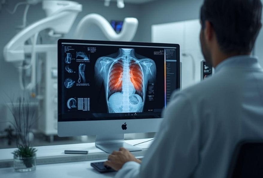
Doctors examine millions of chest scans every year to diagnose pneumonia, tuberculosis, and lung cancer. However, even the most experienced radiologists can miss subtle patterns after reviewing hundreds of images daily. AI chest X-rays are now reshaping medical imaging by acting as a powerful digital assistant that enhances speed and precision.
Recent research, including the 2024 review published in Diagnostics, highlights how deep learning models are improving pulmonary abnormality detection. As a result, hospitals worldwide are adopting AI-driven tools to support clinical decisions.
How AI Chest X-Rays Learn to Detect Disease
Teaching a computer to recognize lung disease is not simple. Engineers train systems using deep learning techniques, particularly Convolutional Neural Networks (CNNs). Initially, the algorithm sees only pixels and grayscale values. Gradually, it begins recognizing structures such as ribs, lung fields, and heart shadows.
Because tumors and infections alter tissue patterns, the AI learns to distinguish abnormal shapes from healthy ones. Consequently, suspicious areas are flagged for radiologists to review.
A major breakthrough came from CheXNet, developed by researchers at Stanford, which demonstrated radiologist-level pneumonia detection using deep learning. This milestone proved that AI chest X-rays could match expert performance under controlled conditions.
How Big Data Drives AI in Medical Imaging
AI systems require massive datasets to perform reliably. Two of the most influential datasets include:
- ChestX-ray14
- MIMIC-CXR
These databases contain hundreds of thousands of annotated chest scans. Because diversity improves fairness, researchers ensure the AI sees images from various age groups and medical conditions. Nevertheless, limitations remain. Some datasets lack demographic balance, which may reduce performance in underrepresented populations.
Therefore, improving dataset diversity has become a priority in medical AI development.
The Role of Computer Vision in AI Chest X-Rays
Computer vision enables machines to interpret visual information. In chest imaging, one essential technique is image segmentation. This process isolates lung regions from bones and soft tissue.
By focusing exclusively on relevant structures, AI chest X-rays improve diagnostic accuracy. In addition, segmentation reduces noise and enhances pattern recognition. As technology advances, these tools continue to refine their predictive capabilities.
Key Challenges in AI-Powered Lung Imaging
Despite progress, challenges persist. One significant issue is the “black box” problem. Deep learning models sometimes produce accurate results without clearly explaining how they reached conclusions.
To address this, engineers are developing explainable AI systems. These tools generate heatmaps that show precisely which lung areas influenced the decision. Consequently, doctors can verify and trust the AI’s reasoning.
Another challenge involves compatibility with older X-ray machines. Since many clinics operate outdated equipment, developers must ensure AI software performs consistently across different hardware systems.
Will AI Chest X-Rays Replace Doctors?
Many fear automation will eliminate medical jobs. However, AI chest X-rays are designed to assist—not replace—radiologists.
AI handles repetitive screening tasks quickly. Meanwhile, doctors interpret complex cases, consider patient history, and make final decisions. As a result, this partnership enhances efficiency while preserving human oversight.
The Future of AI Chest X-Rays
Looking ahead, AI chest X-rays may predict diseases before symptoms appear. Mobile applications for remote diagnostics are already under development. Because healthcare access remains limited in many regions, portable AI tools could dramatically improve early detection rates.
Furthermore, research continues to refine model accuracy and fairness. As these systems evolve, they promise to reduce diagnostic delays and improve survival outcomes worldwide.
AI and the Future of Radiology
AI chest X-rays represent one of the most promising advancements in medical imaging. By combining deep learning, big data, and computer vision, these tools enhance early lung disease detection. Although challenges remain, the collaboration between human expertise and artificial intelligence is shaping a smarter, faster healthcare future.
Additionally, to stay updated with the latest developments in STEM research, visit ENTECH Online.
Reference:
- Parra-Cabrera, G., Jiménez-Delgado, J. J., & Pérez-Cano, F. D. (2026). Artificial Intelligence for Pulmonary Abnormality Detection in Chest X-Ray Imaging: A Detailed Review of Methods, Datasets and Future Directions. Technologies, 14(3), 147. https://doi.org/10.3390/technologies14030147
- Author
- Latest Posts

Kottauppari Venkat Raghava
I am a technology-driven IT graduate with a strong passion for science and innovation. I am fascinated by how innovations like artificial intelligence, robotics, and emerging technologies are shaping our world. I specialize in analyzing complex scientific and technological research and presenting it in a clear, accessible way. My goal is to make science and technology understandable and engaging for students, tech enthusiasts, and professionals alike. By breaking down advanced concepts into simple, insightful narratives, I strive to inspire curiosity, learning, and innovation, helping readers stay informed about the breakthroughs that are shaping the future.


