Innovative Nanosheet Method Revolutionizes Brain Imaging for Multi-scale and Long-Term Studies The human brain is a complex network of billions of neurons that work together to enable higher-order functions such as cognition and behavior. To…
Revolutionary NIRE Method Allows Long-Term and Multi-Scale Brain Imaging
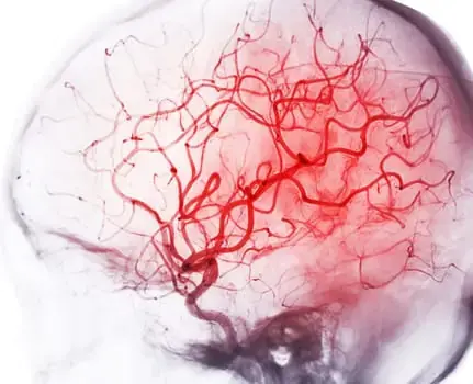
Innovative Nanosheet Method Revolutionizes Brain Imaging for Multi-scale and Long-Term Studies
The human brain is a complex network of billions of neurons that work together to enable higher-order functions such as cognition and behavior. To understand these functions, it is crucial to study how neural activity is coordinated across different regions of the brain. While techniques like functional magnetic resonance imaging (fMRI) can provide insights into brain activity, they have limitations in terms of the amount of information they can show at a given time and area.

However, a team of researchers from the Exploratory Research Center on Life and Living Systems (ExCELLS) and the National Institute for Physiological Sciences (NIPS) has developed a new method for in vivo brain imaging that allows for large-scale and long-term observation of neuronal structures and activities in awake mice. This groundbreaking method, called the “nanosheet incorporated into light-curable resin” (NIRE) method, uses fluoropolymer nanosheets covered with light-curable resin to create larger cranial windows.
Lead author Taiga Takahashi from Tokyo University of Science and ExCELLS explains, “The NIRE method is superior to previous methods because it produces larger cranial windows than previously possible, extending from the parietal cortex to the cerebellum. This is made possible by using a biocompatible nanosheet and transparent light-curable resin that changes from liquid to solid form.”
In this method, light-curable resin is used to fix polyethylene-oxide-coated CYTOP (PEO-CYTOP), a bioinert and transparent nanosheet, onto the surface of the brain. This creates a “window” that fits tightly onto the brain surface, even on highly curved surfaces like the cerebellum. The window maintains its transparency for a long time with minimal mechanical stress, allowing researchers to observe multiple brain regions in living mice.
Takahashi adds, “We also demonstrated that the combination of PEO-CYTOP nanosheets and light-curable resin results in stronger cranial windows with greater transparency for longer periods of time compared to our previous method. This reduces motion artifacts caused by the movements of awake mice, resulting in high-resolution imaging with sub-micrometer resolution to observe the morphology and activity of fine neural structures.”
Corresponding author Tomomi Nemoto from ExCELLS and NIPS explains the significance of this study, “The NIRE method enables imaging for longer periods of more than 6 months with minimal impact on transparency. This will allow for longer-term research on neuroplasticity at various levels, from the network level to the cellular level, as well as during maturation, learning, and neurodegeneration.”
This innovative method has opened up new possibilities for neuroimaging research by providing a powerful tool for studying neural processes that were previously difficult or impossible to observe. With its ability to create large cranial windows and enable long-term imaging, the NIRE method will contribute significantly to advancing our understanding of brain function and behavior.
Source: https://www.excells.orion.ac.jp/en/news/9812
- Author
- Latest Posts

Akanksha Ghodke is a passionate Computer Science graduate from Walchand Institute of Technology, Solapur, with a strong foundation in web development, software engineering, and emerging technologies. She has a deep interest in crafting innovative and impactful digital solutions that enhance user experiences and optimize performance.
With hands-on experience in both front-end and back-end development, Akanksha is proficient in Java, Python, C/C++, ReactJS, NodeJS, WordPress, and database technologies like MySQL, MongoDB, and PostgreSQL. She specializes in designing and optimizing websites, ensuring seamless navigation, responsiveness, and high-performance functionality. Her expertise in UI/UX design, digital marketing, and SEO enables her to build web applications that are not only efficient but also visually appealing and search-engine optimized.
Beyond web development, Akanksha has a strong inclination toward data engineering, cloud computing, and DevOps. She has worked with Docker, Kubernetes, CI/CD pipelines, and data migration, gaining hands-on experience in developing scalable, secure, and automated systems. She continuously explores new trends and technologies, staying up to date with advancements in the tech world.
Her passion for technology extends to her writing, where she simplifies complex topics into informative and engaging content. Some of her favorite subjects include Database Management Systems (DBMS), Java programming, Operating Systems, STEM (Science, Technology, Engineering, and Mathematics), Artificial Intelligence, Blockchain, and emerging innovations in the digital landscape. She actively writes blogs, articles, and guides to share her insights and knowledge with the broader tech community.
Apart from her technical pursuits, Akanksha is an avid reader with a love for biographies and motivational storytelling. She enjoys exploring the lives of inspiring individuals who have made significant contributions in various fields. These stories fuel her determination and drive to make a meaningful impact in the tech industry. In her free time, she also enjoys painting, drawing, and watching web series, balancing her analytical mindset with creativity.
With a strong belief in continuous learning, Akanksha strives to enhance her expertise and contribute to the ever-evolving world of technology. She aspires to develop cutting-edge solutions that empower businesses, improve efficiency, and drive innovation. Her journey in tech is fueled by curiosity, perseverance, and the desire to create a positive impact in the digital world.
- Author
- Latest Posts

Until 2023, Dr. Charudatta S Pathak held multiple academic positions, including lecturer, assistant professor, professor, dean, principal, director, and vice chancellor at public and private universities across India. From 2008 to 2010, he held the position of project lead in the CAE department at a European multinational corporation. Throughout his 28-year professional experience, he observed a requirement for reliable publications aimed at youngsters in grades 8 to 12, specifically for early-stage career planning. He initiated the establishment of ENTECH Digital Magazine, a complimentary periodical released on a monthly basis, accessible via stemconference.in and magzter.com. Teenagers with a keen interest in Science, Technology, Engineering, and Mathematics (STEM) and aspiring to pursue professional paths in these domains can consider reading ENTECH Digital Magazine.


