Discover how super resolution microscopy uses a tiny mirror to image living cells in stunning 3D detail at nanoscale precision.
Scientists Just Built a Microscope That Can See Inside Your Cells Like Never Before
A Mirror Changes Everything in Super Resolution Microscopy
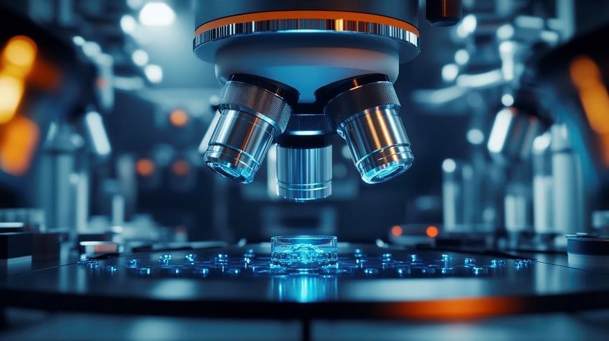
At first, it sounds almost too simple. Scientists added a tiny mirror to an existing microscope. That single change, so far, has produced one of the biggest breakthroughs in biological imaging in years. Researchers at Zhejiang University in China published exciting new findings in Nature Biotechnology on March 31, 2026. Their new technique is called mirror-enhanced 4Pi single-molecule localization microscopy, or me4Pi-SMLM for short (Yu et al., 2026).
What Is Super Resolution Microscopy? Why Should You Care?
Seeing Past the Blur with Super Resolution Microscopy
So what exactly is super resolution microscopy? To explain, regular light microscopes hit a natural limit. They cannot show details smaller than about 200 nanometres. That is roughly 500 times thinner than a human hair. Seeing that, most of the important machinery inside a cell — like proteins, membranes, and tiny organelles — is far smaller than that.
Super resolution microscopy smashes this limit. As a result, scientists can now see structures just 2 to 15 nanometres in size. To illustrate, that is like zooming in on a tennis ball until you can count its individual rubber molecules. Techniques in this field, such as single-molecule localization microscopy (SMLM), work by tracking individual glowing molecules one at a time and combining millions of precise measurements into one ultra-sharp image.
Also Read: Breakthrough in Nuclear Medicine with Perovskite Gamma-Ray Cameras
How the me4Pi-SMLM Mirror Trick Works
A Clever Use of Reflected Light
To enumerate the key idea here: the team placed a tiny protected silver mirror above the biological sample. The laser beam shines up through the sample and then bounces back down off the mirror. With this in mind, the reflected beam creates a pattern of bright and dark bands — called an interference fringe — right at the sample level.
The Result: Near-Isotropic Nanoscale Precision
What’s more, the result is remarkable. The system achieves 2 to 3 nanometre localization precision in all three dimensions — up, down, left, right, forward, and back. To rephrase it: it is equally sharp in every direction. Prior to this, most microscopes saw things clearly only from the side, while depth information remained blurry.
Also Read: Introduction to Optics Physics for High School Students
What Can Scientists Actually See With This?
Tiny Hollow Tubes Inside Cells
At this point, you might wonder: what does all this actually reveal? The researchers imaged several important structures inside cells:
- Microtubules — hollow tube-like structures that give cells their shape. The me4Pi-SMLM system resolved the hollow ring-shaped cross-section of individual microtubules with crystal clarity.
- Nuclear pore complexes — tiny molecular gates in the cell’s nucleus. The system resolved nearly all 32 copies of a protein called Nup96 arranged in an intricate ring, separated by just 10 nanometres.
- Endoplasmic reticulum (ER) — a network of membranes that processes proteins. The system showed sheet-like ER structures just 30 to 50 nanometres thick.
Live Cells and Brain Slices
Seeing that conventional 4Pi systems could not image living cells at all, this is a major leap forward. The me4Pi-SMLM system successfully imaged live cells in real time, tracking ER membrane dynamics over 10 minutes. ER tubules measured 60 to 70 nanometres in diameter. All of a sudden, live-cell nanoscale imaging became possible with a single-objective setup.
What’s more, the team imaged 30-micrometre-thick mouse brain slices — actual tissue, not just cells in a dish. At a depth of 20 micrometres inside the tissue, they still achieved sub-15 nanometre resolution. To point out how big this is: brain tissue is notoriously difficult to image at this scale because it scatters light in unpredictable ways.
Also Read: How Multiscale Aperture Synthesis Imager (MASI) is Advancing Optical Imaging
Why This Matters for STEM Careers: Super Resolution Microscopy
The Upgrade Path Is Surprisingly Simple
While this may be true that high-end equipment upgrades are usually expensive and complicated, this one is different. Almost any existing 3D-SMLM microscope can be upgraded to me4Pi-SMLM. The upgrade requires only a mirror and an affordable piezoelectric actuator. To sum up, this dramatically lowers the barrier for labs around the world. More labs with better tools means faster scientific discovery for everyone.
Career Opportunities This Research Points To
So what kinds of STEM careers connect to research like this? To list some exciting options:
- Optical Engineer — designs and builds advanced imaging systems like me4Pi-SMLM
- Biophysicist — applies physics principles to understand living systems at the molecular scale
- Cell Biologist — uses super-resolution microscopes to study how cells work and malfunction in disease
- Biomedical Engineer — builds tools and instruments for medical research and diagnostics
- Neuroscientist — uses brain-slice imaging techniques to study diseases like Alzheimer’s and Parkinson’s
- Software/Algorithm Developer — writes the AI and image-processing code that turns raw data into sharp images
- Data Scientist — analyses the huge datasets produced by modern microscopes
As a matter of fact, the intersection of optics, biology, computing, and engineering in this single paper shows why STEM careers are so collaborative and exciting. No single person built this — it took a whole team of people with diverse skills. As can be seen in this work, the boundaries between science and engineering are constantly blurring.
Education Pathways to Explore
With this purpose in mind, if you want to work in fields like this, here are some starting points for your grade 11 or grade 12 journey:
- Focus on physics, chemistry, and biology — all three are essential for bioimaging
- Learn basic programming — Python and MATLAB are common tools in imaging labs
- Explore optics — even a basic understanding of how lenses and mirrors work is a huge advantage
- Look into university programs in biomedical engineering, biophysics, or applied physics
For more inspiration, check out ENTECH’s Science section for more articles on cutting-edge discoveries. You can also explore ENTECH’s Engineering section to discover career paths in biomedical and optical engineering.
References:
- Yu, Z., Zheng, B., Zhan, Y., Li, S., Wang, X., Dai, Q., Chen, Y., Wei, Z., Zhao, W., Chen, L., Tang, J., Xia, T., He, L., Liu, C., Wu, X., Yu, X., & Zhang, Y. (2026). Mirror-enhanced 4Pi-SMLM with one objective enables isotropic nanoscale imaging. Nature Biotechnology. https://doi.org/10.1038/s41587-026-03083-7
- Author
- Latest Posts

I am an inspirational writer and emerging science blogger with a deep-rooted passion for understanding and sharing the wonders of the world around us. My journey began with a Bachelor of Science in Chemistry from AKI’s Poona College of Arts, Commerce and Science, where I developed not only a strong foundation in scientific principles but also a mindset that values curiosity, observation, and critical thinking. Studying chemistry has taught me to approach problems with logic, to look beyond the obvious, and to see patterns where others might only see complexity. These experiences have shaped the way I perceive life and influence the way I express my thoughts, both scientifically and creatively.
I have always believed that knowledge is only as powerful as the way it is shared. This belief has guided me to create content that bridges the gap between science and everyday life. I aim to simplify complex scientific ideas so they become approachable, relatable, and engaging for everyone—whether someone has a formal background in science or is simply curious about the world. Science, in my view, is not confined to textbooks and laboratories; it is a way of thinking, understanding, and connecting with the world around us. By making science accessible, I hope to inspire curiosity, ignite learning, and encourage people to see the beauty and relevance of scientific discovery in their daily lives.
Alongside my love for science, I am deeply passionate about motivational writing. I believe words carry the power to transform perspectives, encourage self-reflection, and inspire action. By combining analytical thinking with creativity, I create content that is intellectually stimulating yet emotionally resonant. Whether I am breaking down a chemical concept, exploring a scientific phenomenon, or crafting motivational narratives, my goal is to communicate ideas in a way that is clear, meaningful, and impactful.
Through my work, I strive to empower my readers—not just by helping them understand the science around them, but by inspiring them to grow, learn, and realize their full potential. Every article, post, or story I write reflects my commitment to turning knowledge into inspiration. I want to spark curiosity, encourage self-improvement, and show that learning and personal growth are deeply connected. For me, writing is not just a craft; it is a way to touch minds, connect hearts, and make ideas come alive in a way that truly resonates.

