Doctors and researchers often add fluorescent dyes to cells so they can see live cell structures under a microscope. Those dyes can change how the cell behaves and limit how long you can watch. A new instrument from Stanford reaches about 120-nanometer resolution inside living cells without any labels. Scientists can now watch the cell’s own machines move and interact in real time. This clearer view helps cancer research, drug testing, and studies of how plants interact with microbes. Students in a biology lab once tried to track vesicles with fluorescent tags and saw the signal fade after only a few minutes. The new method avoids that problem.
Images shown here are for illustration purposes only. They are AI-generated and do not necessarily represent the original research.
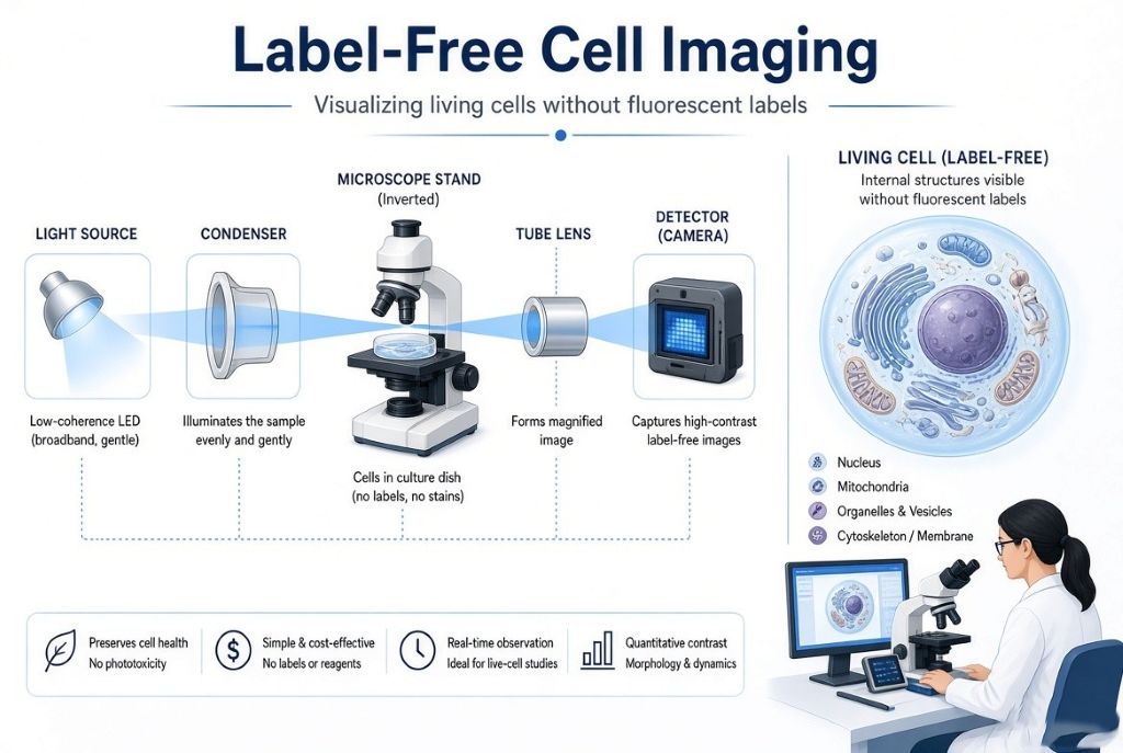
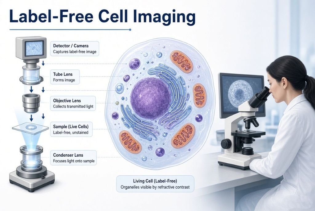
Fig. 1: Stanford’s interferometric method reveals cellular structures in living cells at high resolution without dyes.
How a Live Cell Microscope Works Without Labels
Most high-resolution views of cells rely on fluorescent tags. These tags bind to specific proteins but can alter cell behavior and bleach under light. The Stanford team combined interferometric scattering microscopy with image scanning microscopy. Light scattered by cellular structures interferes with a reference beam. A camera records many small images as the laser scans. Software then reassembles the data into a sharper picture. The result shows the endoplasmic reticulum, mitochondria, vesicles, and actin structures in living cells. Illumination power stays very low, about 0.5 microwatts per spot. This low dose lets researchers image for long periods. Students who have aligned confocal microscopes know how quickly fluorescent signals fade. The label-free approach removes that limit and still delivers high contrast.
Building High-Resolution Live Cell Microscope Images
The instrument uses a custom scanning setup with galvanometric mirrors and a sensitive camera. A 445 nm laser provides the illumination. Polarization is controlled to reduce artifacts. At each scan position, the system captures a microimage. An adaptive pixel-reassignment algorithm reconstructs the final image. The method achieves a lateral resolution of roughly 120 nm. Contrast-to-noise ratio improves about fourfold compared with closed-pinhole confocal versions. Axial sensitivity comes from phase variations, so structures above and below the focus appear with opposite contrast. Live COS-7 cells show clear organelle motion over many frames. The same optical path can accept a second fluorescence laser. Researchers can therefore correlate unlabeled structures with specific labeled molecules when needed. The open-access paper describes the full optical layout and reconstruction steps.
Why Label-Free Imaging Matters for Research
Fluorescent labels give molecular specificity but introduce phototoxicity and limited observation windows. The new technique avoids those problems while still resolving fine structures. Scientists can watch vesicle trafficking, endoplasmic reticulum remodeling, and cytoskeletal changes for extended periods. Drug effects or pathogen interactions become easier to study because the cell remains closer to its natural state. The system also works with plant cells and microbial interactions. Instrument makers and biotech companies need people who can design low-light optical systems and write image-reconstruction code. University labs already use similar scanning architectures, so researchers can adapt the method to existing confocal platforms. Early undergraduate projects often involve measuring point-spread functions or testing reconstruction algorithms on bead samples.
Careers with Live Cell Microscope Technology
Biomedical engineering, biophysics, and optical engineering programs teach the principles behind this instrument. Undergraduate research opportunities often involve aligning optical systems, writing image-analysis code, or testing biological samples. Hospitals, biotech companies, and instrument makers hire people who can operate and improve advanced microscopes. Students who like both biology and physics can start with lab courses in microscopy or image processing. Python skills for analyzing image data are especially useful. Internships frequently begin with practical tasks such as calibrating detectors or processing time-lapse datasets. Later roles include designing new optical modules or developing software for real-time reconstruction. The Stanford work shows that combining established techniques can open new biological questions without requiring entirely new hardware platforms.
How This News Article Was Compiled
The article draws only from the open-access paper in Light: Science & Applications and the official Stanford University research announcement. Both sources were reviewed in full. Technical numbers, methods, and claims come directly from those documents. Career notes reflect standard educational pathways in microscopy and biophysics.
Küppers, M., & Moerner, W. E. (2026). Interferometric Image Scanning Microscopy for label-free imaging at 120 nm lateral resolution inside live cells. Light: Science & Applications, 15. https://doi.org/10.1038/s41377-026-02210-y
Mahi Shitut is a Science Communicator at ENTECH STEM Magazine, with a BSc in Physics from Fergusson College, Pune, and an ongoing MSc in Mathematics at the Indian Institute of Science Education and Research (IISER), Kolkata. Her areas of interest include theoretical physics, pure mathematics, astronomy, space science, set theory, asymptotic series, and science education. She completed the National Initiative in Undergraduate Science (NIUS) Physics Programme at the Homi Bhabha Centre for Science Education, TIFR, Mumbai, where she carried out a 1.5-year research project at the UM-DAE Centre for Excellence in Basic Sciences. She was also a top scorer in the Madhava Mathematics Competition and was selected for the Madhava Nurture Camp at IIT Bombay, and she attended the IWM Winter School for Young Women in Mathematics.
Mahi specializes in explaining advanced physics and mathematics in simple, clear, and easy-to-understand language for teenagers and school students. Drawing on everyday examples and practical analogies, she helps readers make sense of ideas like infinity, space missions, telescopes, and how the universe works, without losing scientific accuracy. Her skill comes from hands-on experience. She has explained models of Chandrayaan-3 to more than 3,000 visitors at IUCAA’s National Science Day open day, volunteered at SPPU’s National Space Day celebrations, presented at Fergusson College’s Astro Club Poster Exhibitions, and delivered undergraduate lectures on countability and uncountability, the infinite hotel paradox, and asymptotic series through the Synectics Math Club. With earlier experience in editorial work and content strategy, including editing a newsletter and writing for digital audiences, she aims through ENTECH STEM Magazine to bridge the gap between advanced science and public understanding, making physics and mathematics engaging, accessible, and relevant for young minds.
Ishika Tawar is a biotechnology researcher and science enthusiast with a curiosity for understanding how life works at the molecular and cellular level. That curiosity started somewhere around school, wandered through a lot of unrelated corners of science and physics, and eventually settled on something she actually wants to spend her life on: aging and longevity. She supports transhumanism and believes that science and technology should be explored seriously as tools to overcome some of the biological limitations of the human body.
Ishika holds a Master's degree in Biotechnology and has experience in both wet-lab and computational research. At ACTREC, she worked on a project studying the interaction of the TG2 protein with the ribosome, where she gained experience in cloning, site-directed mutagenesis, transformation, and gene knockdown. She was also involved in studying ribosomal heterogeneity using R and publicly available single-cell RNA sequencing datasets from different cancers. As a Research Assistant at SafeWright, she worked on client-based projects involving microbial culturing, biochemical tests, and analysis, along with literature research, report writing, and preparation of laboratory protocols. She has also worked with Python-based analysis to study differential DNA methylation in breast cancer.
Her current areas of interest include epigenetics, genomics, gene therapy, stem cell biology, computational biology, and the use of AI in biological research. She is particularly interested in how these areas can contribute to understanding aging and developing ways to extend healthy human lifespan. She has explored this interest through research projects and hackathons focused on AI, aging, and longevity. She enjoys following ideas across biology, technology and physics, and she believes in evidence, questioning ideas and staying curious about things that are not yet understood.
Outside research, Ishika enjoys reading and talking about science and scientific ideas. She loves travelling and is especially fond of mountains. She also enjoys science communication and hopes to make scientific topics easier to understand without making them inaccurate or oversimplified.