A newly identified protein controls the intermembrane space between mitochondrial membranes — a discovery that reframes how our cells tune their power plants.
SPRIZ: The Molecular Pillar That Sets Mitochondria’s Inner Spacing
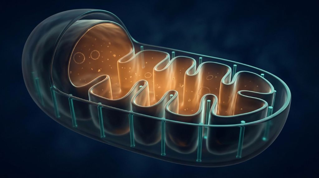
For decades, scientists believed the narrow gap inside mitochondria was a passive feature—merely a consequence of the organelle’s double-membraned structure. But new research has uncovered SPRIZ, a protein that acts as a molecular pillar and actively controls the spacing between the mitochondrial intermembranes. This breakthrough not only reveals a hidden layer of mitochondrial architecture, but also hints at how cells fine-tune their energy production and signaling.
A Molecular Pillar Inside the Mitochondrial Intermembrane Space
Every time your muscles contract as you’re running, billions of little engines (mitochondria) within your cells are burning fuel to get you going. In fact, each of those little engines is a double-membrane structure with a very narrow space between the two membranes, known as the intermembrane space. As a result, ions, signalling molecules, and the various building blocks all pass through that space as they go in and out. For a long time, scientists thought the intermembrane space formed simply because the two membranes came together. However, a team at the University of Ottawa have now discovered a protein that actively maintains the two membranes at a fixed distance from each other. They’ve named it SPRIZ. Consequently, their work in Science Advances shows that the space within the mitochondrion is not just a passive feature; the cell can actively control it.
Key Takeaways: SPRIZ residing in the Mitochondrial Intermembrane Space
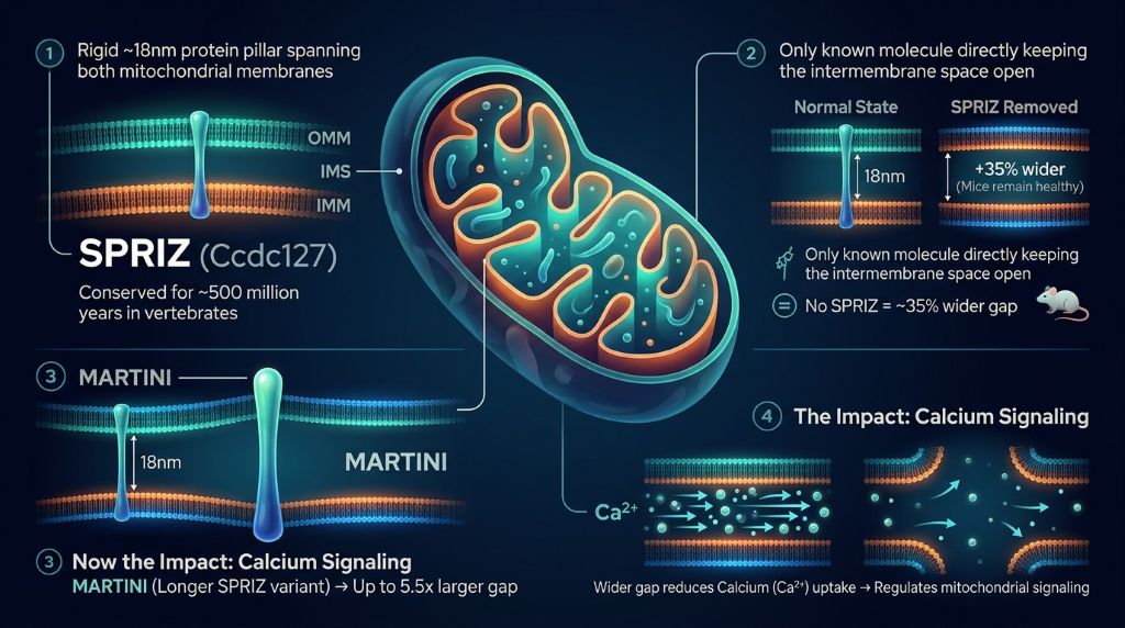
Scientists have recently discovered SPRIZ, a rigid ~18nm protein pillar that spans the two mitochondrial membranes. Notably, this breakthrough reveals not only the first known molecule to control this compartment, but also how tiny shifts in spacing can reshape how mitochondria signal within our cells
The Inner Architecture of the Mitochondrial Intermembrane Space
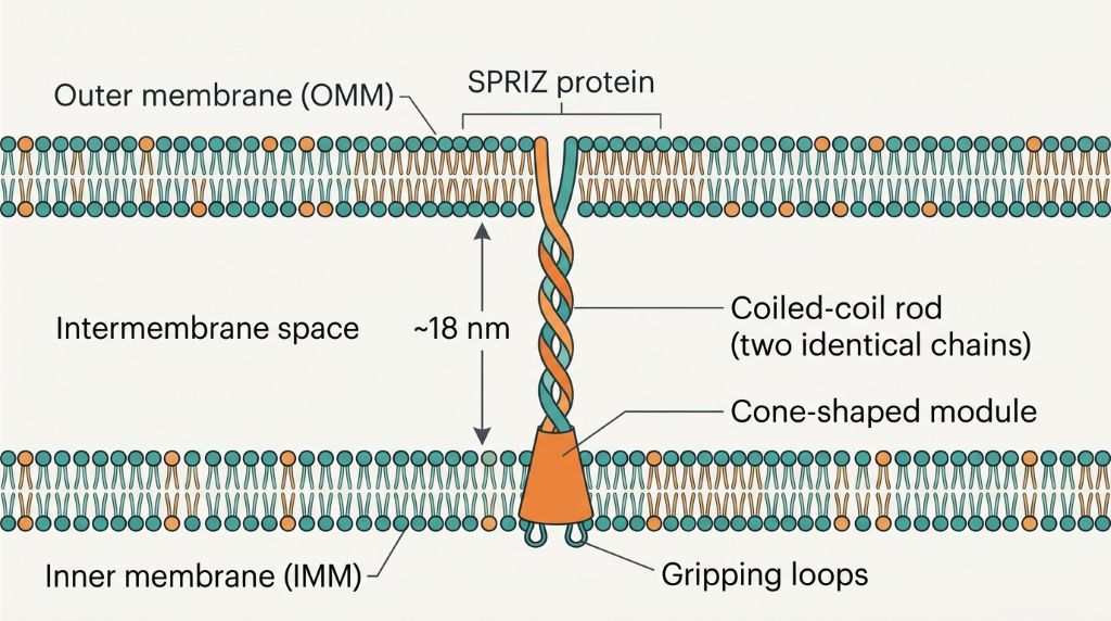
To understand SPRIZ, imagine a double-walled tent with a very narrow gap between the two walls. The outer mitochondrial membrane (OMM) is the first wall. Meanwhile, the inner mitochondrial membrane (IMM) is the second wall. The space between them is the mitochondrial intermembrane space (IMS). Ions, reactive oxygen species, and metabolites must cross the gap to access the inner matrix, where energy production occurs. Until now, it was not known whether the width of that gap is genetically determined.
Also read: Skeletal System
What SPRIZ Actually Looks Like
To uncover the protein in mitochondrial intermembrane space called is a classic tether doing something different (holding the room at a fixed height).
A Feature That Arrived With Vertebrates
SPRIZ was not present in all living things. Remarkably, among 605 high-quality genomes from 31 animal groups, SPRIZ appeared exclusively in deuterostomes, including vertebrates. It was absent from sponges, worms, flies, yeast, and plants. Evolutionary evidence points to an origin at the base of the bilaterians, with near-complete retention throughout the deuterostomes and, in particular, the vertebrates. Over roughly 500 million years, strong purifying selection has maintained the length of the rod and the membrane-gripping loops. The fact that this architecture has been so conserved suggests a conserved function. As animals got larger and developed specialised tissues, their mitochondria would have required much more precise control of the interior space.
For anyone else looking to get a handle on the wider biology of these organelles, our comparison of chloroplasts and mitochondria provides an accessible starting point for how these two energy organelles differ. The full study is open access at Science Advances.
Why This Question Was So Difficult to Answer
Interestingly, it all started with a puzzle. Previously, researchers found no molecule setting intermembrane space dimensions in yeast, worms, or flies; thus, indirect organisation seemed likely. In contrast, vertebrate mitochondria were a little different. Consequently, vertebrates contain more proteins and perform specialised tasks, indicating they may have evolved a system to set the intermembrane space.
The Obstacles the Team Faced
Confirming that notion was by no means easy. The protein in question was entirely uncharacterised and had no known function. You had to work out where it was, what it looked like and what it did from the ground up. The gap it regulates is only a few nanometres wide, so detecting any change required electron tomography and painstaking 3D reconstruction. The protein itself is a very subtle player. Mice lacking SPRIZ were perfectly healthy and viable, developed normally, had normal hearts and livers, and had intact cristae. The only thing you could see was a very small increase in the intermembrane space (a 35% increase to be precise). So you couldn’t rely on any obvious phenotype. You had to look at the organelle itself, one membrane pair at a time.
Building a Tool to Test the Idea Directly
To prove that SPRIZ is genuinely responsible for the spacing, they made a sharper version. They took a segment of the coiled coil and duplicated it to create an artificial longer protein which they called MARTINI (Mitochondrial Artificial Inhibitor of the IMS – the name is a bit of a misnomer as it’s actually an increaser). When delivered into mouse liver cells, SPRIZ increased the IMS area by about a factor of 3, whereas MARTINI, being longer, increased it by about a factor of 5.5. Both effects were highly significant and graded (the longer the rod, the greater the expansion). A mutant SPRIZ in which the membrane-gripping loops were deleted could not expand the IMS. The clear length-dependent, graded response is very strong evidence that it’s the protein itself (not some secondary effect) that is responsible for the expansion.
How This Mitochondrial Intermembrane Space Shapes Calcium Signalling
A bigger room changes what goes on in it. Calcium is the clearest example. Calcium must move from the pores in the outer membrane, across the mitochondrial intermembrane space, and then to a transporter on the inner membrane. A wider gap means a longer distance to travel, and the calcium gets diluted along the way. As a result, mitochondria expressing SPRIZ or MARTINI took up calcium more slowly. The cells lacking SPRIZ did the reverse. The calcium handling machinery itself was unchanged. The difference was entirely a matter of geometry, not a different pump.
A Tunable Parameter, Not a Fixed Setting
The findings are profound: they completely reframe the intermembrane space. Rather than a passive consequence of membrane shape, it’s now clearly a genetically determined and tunable parameter of the mitochondrion. Different tissues express different levels of SPRIZ, and the brain has by far the highest levels. This implies that different cell types could up- or downregulate spacing to meet their own energy and signalling requirements. Given that many organellar processes depend on diffusion and the establishment of local concentration gradients in the intermembrane space, a change in its width could cascade into effects on metabolite exchange, ROS signalling, protein import, and lipid transfer.
One point to note is that calcium uptake in this study serves as a readout of intermembrane space geometry rather than the protein’s sole biological function. The authors make it clear that SPRIZ is expected to affect any process dependent on diffusion or local gradients across the gap. That constraint keeps the result precise; the spacing is changing, and the calcium uptake reports on that change. For other open access work on this topic, see the review of the mitochondrial intermembrane space.
Explainer: The Mitochondrial Intermembrane Space in Plain Language
Think of a mitochondrion as a water balloon inside a slightly bigger balloon. The inner water balloon contains the fuel, and it folds into shelves (the cristae). The outer balloon is the boundary of the mitochondrion with the rest of the cell. A thin layer of water between the two balloons (the mitochondrial intermembrane space) isn’t empty. Protons, calcium ions, and lots of proteins fill that space, and it acts as a corridor for them to move from the cell to the mitochondrion’s inner core.
SPRIZ is the spacer that sets the hallway’s thickness. Two identical SPRIZ chains are twisted together to form a single stiff rod. One end is bolted into the outer membrane, and the other end extends across to grip the inner membrane. The rod doesn’t flex much, so it effectively pins the two membranes a fixed distance apart. Add more SPRIZ and the hallway widens. Use the MARTINI trick to shorten the rod, and the hallway will widen even more. Remove the loops that are grabbing the inner membrane, and the hallway will snap back to its normal width.
This is why the discovery matters. Hallway width determines how quickly people (in this case, ions and signals) can pass through it. When I teach this in the classroom, I often use the analogy of dragging a heavily loaded cart through a corridor. If you widen the corridor, the task changes even though the cart and the load are the same. SPRIZ does the same thing for calcium within the cell. It alters the hallway, not the cargo.
What This Means for Science and Medicine
The immediate value of SPRIZ is conceptual: it provides biologists with the first dedicated molecular system known to set the distance between mitochondrial intermembrane space. That fills a gap in our understanding of how these organelles assemble and are regulated. The study also gives other labs a reusable MARTINI tool that can expand space on demand and let them investigate how a larger compartment affects various cellular processes.
The medical relevance is more remote but certainly there. Mitochondria often misfire in diseases like Parkinson’s and energy-deficient conditions; consequently, SPRIZ defects could plausibly influence disease risk. The authors are very cautious about this and don’t claim any such involvement in disease. What they do is open a whole new avenue of investigation, where the shape of the mitochondrion is as important as its chemistry.
This brings us back to the bigger picture. For a look at how this kind of structural control could one day be applied in a clinical context, see our piece on the promise of mitochondrial gene editing and the primary paper in the Science Advances article for the full methods and data.
Conclusion
For a long time, biology just regarded the mitochondrial intermembrane space as a passive consequence of other things. The discovery of SPRIZ completely changes that. It’s a protein that is essentially a pillar, highly conserved for half a billion years, and its job is to keep the two membranes a set distance apart. The protein’s length determines the width of the intermembrane space, and that width in turn dictates calcium signalling within the cell. It’s like the cell deliberately building a small room. It also reminds us that dimensions matter as much as chemistry in determining what a cell can do.
References
Edwards, R., Gerlich, S., Tokatlidis, K., & Lu, H. (2021). The mitochondrial intermembrane space: The most constricted mitochondrial sub-compartment, the verge of discovery. PMC. https://pmc.ncbi.nlm.nih.gov/articles/PMC8061763/
Shulinski, R., Loban, R., Farhat, D., Cartes-Saavedra, B., Magda, M. J., Kotchetkov, P., Caminiti, V., David, L., Wolf, Y. I., O’Dwyer, C., Fullerton, M., Burelle, Y., Lee, J.-Y. (E.), Raimondi, A., Koonin, E. V., Hajnóczky, G., Harper, M.-E., & Pellegrini, L. (2026). SPRIZ (CCDC127) protein conserved in deuterostome animals is a rigid OMM–IMM tether that locally sets mitochondrial IMS volume. Science Advances, 12(42), eaef3522. https://doi.org/10.1126/sciadv.aef3522
University of Ottawa. (2026, October). uOttawa scientists uncover the molecular pillars that shape our cells’ powerpacks [Press release]. Faculty of Medicine. https://www.uottawa.ca/faculty-medicine/news-all/uottawa-scientists-uncover-molecular-pillars-shape-our-cells-powerpacks
- Author
- Latest Posts

Sailee Pakhare holds a Master’s degree in Biotechnology, with a research background spanning microbiology, molecular biology, and disease biology. Her postgraduate dissertation examined the antimicrobial potential of bioactive peptides from lactic acid bacteria, and her research experience includes molecular identification of bacterial isolates (with sequencing data submitted to NCBI) and work as a Research Assistant at SafeWright Laboratory. She also completed a summer internship at IIT Hyderabad studying circadian rhythms and disease biology using Drosophila melanogaster as a model organism.
Her work has been recognized with the Best Poster award at the 3rd International Conference on Innovations in Biotechnology Research for Sustainable Bioresources and Bioeconomy (2025), and she has contributed to a book chapter on the role of AI and machine learning in climate change.
Sailee is also a co-founder of LifeXtension, a community focused on longevity, ageing, and health science awareness in India. An interdisciplinary thinker, she explores questions ranging from disease biology to space and the origins of life, approaching science as a way of asking questions rather than a fixed body of facts. As a science communicator and researcher, she is committed to translating complex research into writing that is clear, accurate, and engaging.