Bones have a unique structure, being made of living tissue that is both strong and lightweight. The hard and dense outer layer gives bones...
Skeletal System: Understanding Bones, Joints, and Movement
Estimated reading time: 8 minutes
Your skeleton is not a dry, dead scaffold. It is a living, dynamic organ system that rebuilds itself every day. Think of it as your body’s personal architect. It gives you shape and allows you to stand tall. Without it, you would be a shapeless pile on the floor. Every movement you make, from blinking to running, depends on your bones and the joints that connect them. This framework also protects your most vital organs, like your brain and heart. Surprisingly, your bones even help your body regulate calcium levels. So, the skeleton does much more than just hold you up. Let us explore how this incredible system works, grows, and stays healthy. You will learn about its structure, its functions, and how to keep it strong for life.
Key Takeaways
- Common disorders like osteoporosis and fractures are often preventable with proper nutrition and exercise.
- Your skeleton has five critical jobs: support, protection, movement, mineral storage, and blood cell production.
- Bones come in five shapes: long, short, flat, irregular, and sesamoid.
- The skeleton splits into two main parts: the axial (head and trunk) and the appendicular (limbs and girdles).
- Bone tissue is either compact (dense) or spongy (porous), with cells that build and break down bone.
- Joints are classified by their structure and the range of movement they allow.
Why the Skeletal System Matters — 5 Essential Functions

The skeletal system serves as the body’s structural cornerstone and performs several essential functions. First and foremost, its primary role is support, as it provides a strong framework that holds soft tissues in place and gives the body its shape. Without this rigid framework, the body would not be able to maintain an upright posture or perform everyday activities such as standing and sitting. In addition to providing support, the skeletal system plays a crucial role in protection. For example, the skull surrounds and shields the brain, while the rib cage protects vital organs such as the heart and lungs. Furthermore, bones contribute to movement by acting as levers for muscles.
Bone Classification — Long, Short, Flat, Irregular, Sesamoid
Human bones are classified into five distinct shapes, and each type is adapted to perform a specific function. First, long bones are characterised by being longer than they are wide. Because of their structure, they primarily function as levers that support movement. For example, the femur, or thigh bone, and the humerus, or upper arm bone, are both long bones. In contrast, short bones are approximately cube-shaped and are mainly responsible for providing stability while allowing limited movement. For instance, the carpals in the wrist and the tarsals in the ankle are typical examples of short bones. Furthermore, while long bones are particularly important for movement, short bones provide strength and stability in areas that require controlled motion. Thus, the distinct shapes of bones are closely related to their specific roles within the skeletal system.
List of Bone Types with Examples
- Long bones: Femur, tibia, humerus, radius
- Short bones: Carpals, tarsals
- Flat bones: Skull bones, ribs, scapulae
- Irregular bones: Vertebrae, sacrum, mandible
- Sesamoid bones: Patella, pisiform
Bone shape is directly linked to function. Long bones maximize movement. Flat bones maximize protection. Irregular bones handle complex forces.
Axial vs. Appendicular Skeleton — A Clear Breakdown
The human skeleton is divided into two main divisions, each serving important structural and functional roles. First, the axial skeleton forms the central axis of the body and provides essential support and protection. Specifically, it includes the skull, vertebral column, and rib cage, comprising approximately 80 bones in adults. In particular, the axial skeleton protects vital organs and supports the body’s overall structure. For example, the vertebral column surrounds and protects the spinal cord, while the rib cage shields the heart and lungs. Furthermore, the axial skeleton provides important attachment points for muscles and for components of the appendicular skeleton. In this way, it helps connect the central framework of the body with the limbs and associated structures. Thus, through its combined roles in support, protection, and structural connection, the axial skeleton forms the essential central framework of the human body.
Bone Histology — Compact vs. Spongy Bone
Your bones are not completely solid throughout; instead, they are composed of different types of bone tissue. Specifically, bone tissue can be broadly classified into compact bone and spongy bone, each serving important structural functions. First, compact bone forms the hard, dense outer layer of most bones. Because of its dense structure, it provides strength, supports the body, and helps resist mechanical stress. Furthermore, when viewed under a microscope, compact bone has a distinctive cylindrical organisation. These cylindrical structures, known as osteons or Haversian systems, are the basic structural units of compact bone. Within each osteon, there is a central canal that contains important blood vessels and nerves. Surrounding this central canal, concentric layers, or rings, of bone matrix are arranged in a circular pattern.
| Feature | Compact Bone | Spongy Bone |
|---|---|---|
| Structure | Dense, solid | Porous, lattice-like |
| Location | Outer layer of bones | Inner layer, ends of bones |
| Units | Osteons (Haversian systems) | Trabeculae |
| Function | Provides strength | Reduces weight, houses marrow |
| Cell activity | Primarily osteocytes | Higher osteoblast activity |
Bone Remodelling & Growth — From Cartilage to Calcium Regulation
Bones continue to grow and remodel throughout life through several coordinated biological processes. Among these processes, endochondral ossification is responsible for forming most bones in the human body. Initially, this process begins with a temporary cartilage model that provides a framework for bone development. Over time, specialized cells called osteoblasts gradually replace the cartilage with bone tissue. In particular, this process is especially important at the epiphyseal plates, or growth plates, located near the ends of long bones. As a child grows, new cartilage is continuously produced on one side of the growth plate. Meanwhile, cartilage on the opposite side is gradually replaced by bone through ossification.
Joint Classification — Fibrous, Cartilaginous, Synovial
Joints connect two or more bones and allow the skeleton to provide both stability and movement. Generally, scientists classify joints according to their structure and range of movement. First, fibrous joints connect bones with dense connective tissue and allow little or no movement. For example, the sutures of the skull firmly connect the skull bones and protect the brain. In contrast, cartilaginous joints connect bones with cartilage and permit limited movement. For instance, the joints between the vertebrae contain intervertebral discs that provide flexibility and help absorb mechanical shock. Furthermore, these joints allow the spine to bend and move while maintaining stability. Therefore, fibrous and cartilaginous joints demonstrate how the body adapts joint structure to meet different functional demands. Overall, by combining strong connections with controlled flexibility, joints help the skeletal system support the body and facilitate movement.
Major Bones — An Identification Guide
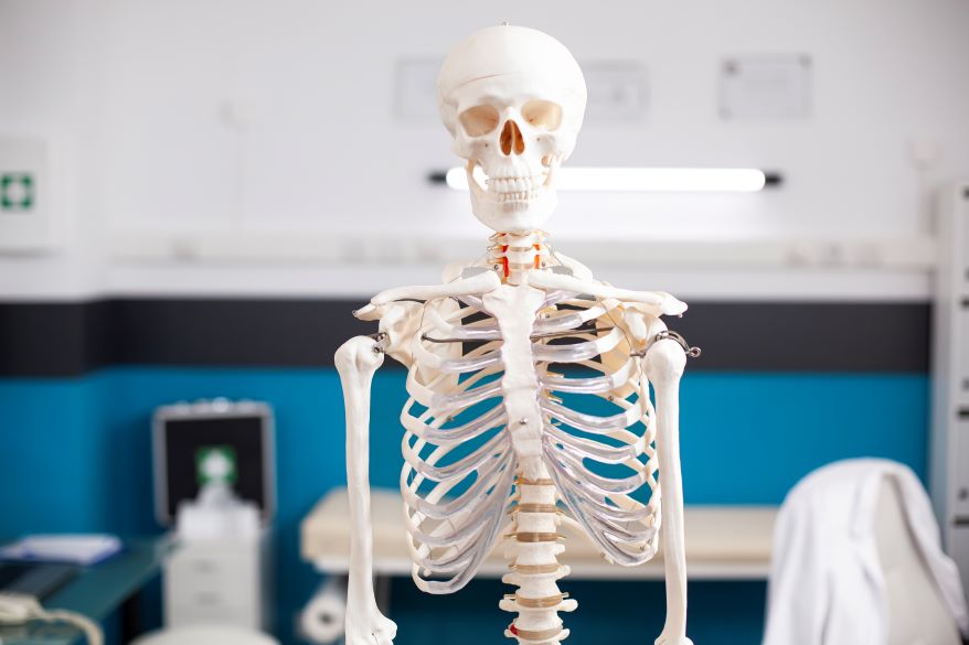
Learning the major bones builds your anatomical knowledge. Start with the skull. It has 22 bones, including the cranium and facial bones. The mandible is your lower jaw. Move down to the vertebral column. It consists of 33 vertebrae. These are divided into cervical, thoracic, lumbar, sacral, and coccygeal regions. The sternum sits in the centre of your chest. Your ribs attach to it. You have 12 pairs of ribs.
Common Skeletal Disorders — From Osteoporosis to Fractures
Several disorders affect the skeletal system. Osteoporosis makes bones weak and brittle. It occurs when bone resorption outpaces bone formation. This condition often affects older adults. Women are at higher risk after menopause. Prevention includes adequate calcium and weight-bearing exercise. Arthritis is joint inflammation. Osteoarthritis is a wear-and-tear condition. It damages cartilage. Rheumatoid arthritis is an autoimmune disease. It attacks the joint lining. Both cause pain and stiffness.
Bone Health for Students — Calcium, Vitamin D, and Exercise
Building strong bones starts early in life and requires consistent attention to nutrition and healthy habits. In particular, peak bone mass is generally achieved around the age of 30; therefore, the choices students make during adolescence and early adulthood can have lasting effects on bone health. First, calcium plays a critical role in building and maintaining strong bones. For example, good sources of calcium include dairy products, leafy green vegetables, and fortified foods. Furthermore, teenagers should aim for approximately 1,000 mg of calcium per day, depending on individual nutritional needs. In addition, vitamin D is essential because it helps the body absorb calcium efficiently.
Frequently Asked Questions (FAQs)
What is the skeletal system?
The skeletal system is the framework of bones as well as cartilage that supports and protects the body. It provides structure, enables movement, produces blood cells, and stores minerals such as calcium and phosphorus.
How many bones are in the human body?
An adult human typically has 206 bones. However, this number can vary slightly due to anatomical differences such as extra ribs or vertebrae.
What are the main functions of bones?
Bones serve several key functions: they provide structural support, protect vital organs, facilitate movement by acting as levers for muscles, produce blood cells in the bone marrow, and also store essential minerals.
How do bones grow and repair themselves?
Bones grow through a process called ossification, where cartilage is gradually replaced by bone tissue. They also continuously remodel by breaking down old bone and forming new bone, which helps repair fractures and maintain strength.
What is bone marrow, and what role does it play?
Bone marrow is the soft tissue found inside bones. It produces red blood cells, white blood cells, and platelets, which are essential for carrying oxygen, fighting infections, and also clotting blood.
Can bones change throughout a person’s life?
Yes, bones change throughout life. They grow in size during childhood and adolescence, and continue to remodel in adulthood. Bone density can decrease with age, especially in conditions like osteoporosis.
What minerals are stored in bones?
Bones primarily store calcium and phosphorus, which are vital for maintaining bone strength and supporting various bodily functions
References
Gan, D., Qin, L., & Xiao, G. (2026). Interorgan communications in skeletal pathophysiology: From molecular pathways to multidisciplinary therapies. Research, 9, Article 1319. https://doi.org/10.34133/research.1319
Maryum, A., Shaukat, A., Yousaf, E., Haque, A., Yusuf, S., Haque, S. U., Ali, H., Jawed, S., & Akram, M. U. (2026). A feature-efficient dual-task machine learning framework for predicting bone mineral density and osteoporosis stratification in resource-constrained environments. PLOS ONE, 21(7), e0354038. https://doi.org/10.1371/journal.pone.0354038
- Author
- Latest Posts

I am a Master’s qualified in Science with focus on Zoology and Entomology and am a passionate, curiosity-driven science enthusiast aiming to transform complex scientific ideas in a manner that are easy to understand hence, educate, and inspire.
I have an experience of working on the projects in the field of Entomology which has provided me hands-on experience with various research techniques. Besides, I have also gained experience of lab work as well as developed communication skills during the course of my bachelors and masters.
With a strong foundation in life sciences and a growing interest in Medical and Biological research, I have cultivated a writing style that merges scientific facts with creativity.
My experience in teaching has also taught me to develop an understanding of what are the interests of people and how to communicate them effectively.
I am committed to lifelong learning, actively engaging in Interdisciplinary learning, so as to broaden my perspective.
- Author
- Latest Posts

I completed my Master of Science from the University of Allahabad in 2017 with a strong academic background in life sciences and chemistry. My specialization included Molecular Biology, Microbiology, Genetics, Plant Breeding, Phycology, Paleobotany, and Bioinformatics, along with Organic Chemistry, Inorganic Chemistry, and Physical Chemistry. This multidisciplinary training provided me with a comprehensive understanding of biological systems and analytical scientific approaches.
After completing my postgraduate studies, I gained valuable professional experience in both the education and social development sectors. I worked at A.M. Oxford Public School, where I was actively involved in guiding students toward academic excellence. In this role, I focused on creating an engaging learning environment, encouraging critical thinking, and nurturing students’ curiosity for scientific learning. My experience as an educator strengthened my communication, mentoring, and classroom management skills.
In addition to my teaching experience, I worked with Jeevan Jagriti Foundation, where I contributed to community-based initiatives aimed at improving educational access and awareness among underprivileged sections of society. Through this work, I participated in programs designed to support social development and promote the value of education in marginalized communities.
These professional experiences helped me develop strong interpersonal, leadership, and organizational skills while reinforcing my commitment to education and community service.