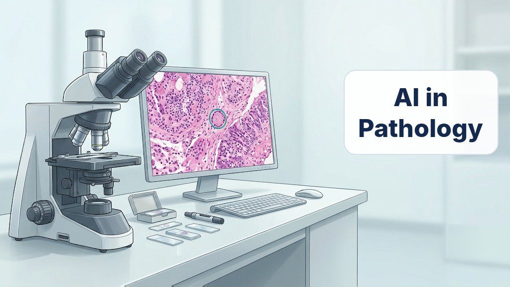
Many health stories include the same quiet sentence. The doctor says a sample has gone to the laboratory. There, a specialist called a pathologist studies thin slices of tissue under a microscope. Their report often decides what treatment, if any, comes next. Screening programs, however, now send far more samples than before. As a result, laboratories face a growing pile of glass slides. AI in pathology is one proposed answer to that pile. Software, in short, could sort the slides so experts focus on the worrying ones. One recent example is a study led by Tsinghua University on cervical tissue. This explainer therefore covers the problem, the method and what might change.
Cervical cancer, in particular, shows the problem clearly. According to the World Health Organization, it is the fourth most common cancer in women. In addition, the agency counted about 660,000 new cases and 350,000 deaths in 2022. Even so, the disease is largely preventable through vaccination, screening and early treatment. For that reason, its 2030 targets include screening 70% of women. More screening is good news, but it creates more follow-up work. When a screening result looks abnormal, doctors may take a small tissue sample. That step is called a biopsy, and the US National Cancer Institute explains such follow-up. Every one of those samples then needs an expert reader. AI in pathology targets exactly this reading step.
The Human Limits of the Microscope
Reading a slide is, above all, skilled and careful work. The pathologist must judge whether cells look normal, mildly changed or dangerous. A mistake in either direction matters, because treatment follows the report. To illustrate, calling a mild change serious can lead to unnecessary treatment. Missing a serious change, on the other hand, can delay care. Even experienced specialists, moreover, sometimes disagree on borderline cases. What is more, the work is slow and places a heavy load on pathologists. The study’s authors describe this workload as complex and labor-intensive. Our primer on pathology science explains the laboratory’s wider role. So the question is simple: can software safely share the reading?
How AI in Pathology Learns to Read Slides
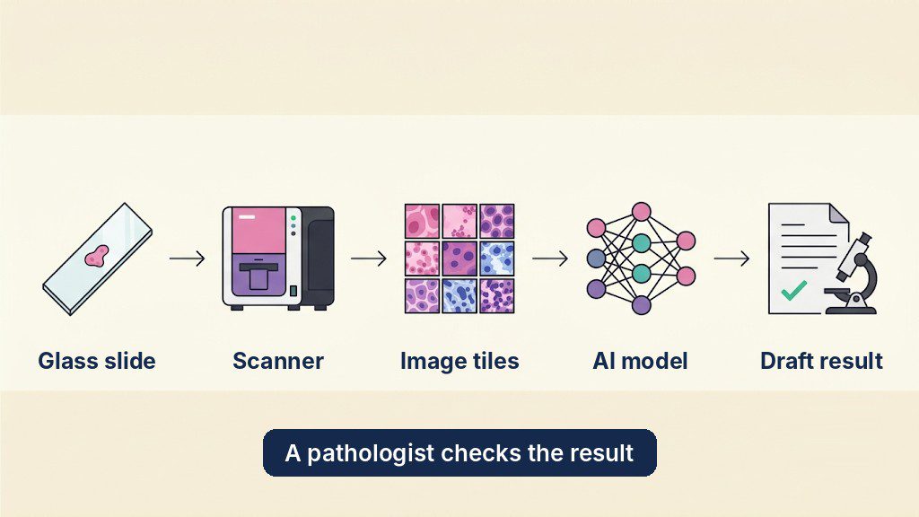
To begin with, glass must be turned into data. A scanner photographs each slide at high magnification, which creates a huge digital image. Because that image is too large to process at once, software cuts it into small tiles. A computer model studies the tiles and combines its findings for the whole slide. Early systems, however, learned only from tiles that experts had labeled by hand. That approach works, but labeling is slow and expensive. It also tends to produce narrow tools that handle one task each. Consequently, researchers looked for ways to learn from unlabeled images. Hospital archives, after all, hold far more unlabeled slides than labeled ones. That is where modern AI in pathology begins.
Explainer: Self-Supervised Learning and Foundation Models
Self-supervised learning, in short, lets a model teach itself from raw images. For example, the software may hide part of a tile and guess the rest. It may also compare two altered views of one tile and learn they match. As a result, the model recognizes textures, shapes and cell patterns after millions of puzzles. Nobody had to label those images first. The result is called a foundation model, because other tools build on it. Engineers then add small labeled sets to tune it for specific tasks. In fact, several general foundation models for pathology already cover many organs. The study’s authors argue, though, that such generalists can miss organ-specific detail. Hence the growing interest in specialist models for AI in pathology.
Fig. 1: From Glass Slide to Draft Result
Explainer: Sensitivity and Specificity
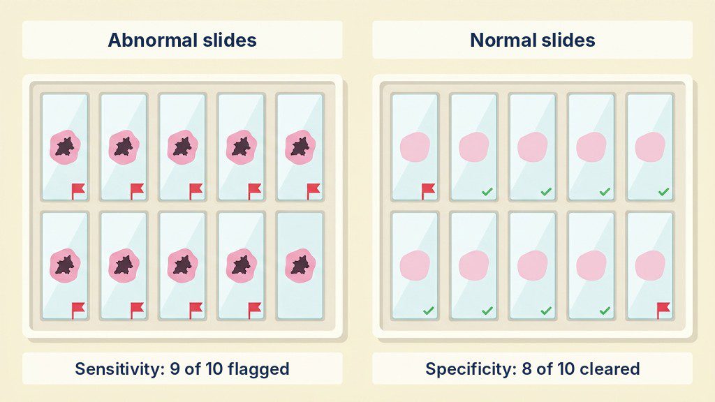
Two numbers describe how well any screening tool performs. Sensitivity is the share of truly abnormal samples that the tool flags. Specificity, in contrast, is the share of normal samples that it correctly clears. For instance, suppose 100 slides contain a lesion, meaning an area of abnormal tissue. A tool with 99% sensitivity would flag 99 of them. Now suppose another 100 slides are normal. With 85% specificity, the tool would clear 85 and wrongly flag 15. Those 15 false alarms then go to a person for checking. Designers must choose which kind of error matters more. In screening, missing disease is usually worse, so tools lean toward high sensitivity.
Fig. 2: Two Numbers Behind Every Screening Tool
A Specialist Pathology AI Built for One Organ
In November 2026, a team reported a specialist system for cervical tissue. Researchers at Tsinghua University’s Shenzhen graduate school led the work together with several hospitals. Partners included the University of Oxford, and the paper appeared in Nature Communications. The system, named CerS-Path, was built in stages. First, a vision model taught itself from about 190 million tiles. Those tiles came from roughly 115,000 scanned cervical slides. After that, the team paired images with text, using about 2.5 million examples. That step, together with a language model, lets the system describe what it sees. Finally, engineers tuned it for 30 tasks grouped into eight functions. These include screening, grading, measuring tumors and drafting reports.
What the Hospital Tests Showed
How did the specialist compare with a generalist? Overall, it beat a leading general model on 26 of 30 benchmark tasks. The average gain was 3.66 percentage points. Then the team ran the system on a working digital pathology platform. From August 2024 to May 2025, it faced slides from five clinical institutions. Likewise, a public dataset was included, with varied stains and scanners. For finding any lesion, sensitivity stayed above 99.0% at every site. Specificity, by comparison, stayed above 84.4%. For lesions that need treatment, sensitivity exceeded 98.6% and specificity 88.4%. In other words, the system rarely missed disease but flagged some normal slides.
How AI in Pathology Could Disrupt Health Care Business
Why does this matter beyond one disease? For the most part, a pathologist today reads every slide from start to finish. With reliable triage, AI in pathology could instead rank slides by urgency before human review. For now, diagnostic laboratories and digital pathology firms can study such specialist models. Within about five years, they might cut backlogs, provided that trials and regulators agree. The US Food and Drug Administration, for example, reviews AI-enabled tools as medical devices. In the long run, similar models could follow for other organs. Pathology work might then shift toward supervising software and solving hard cases. Start-ups may also find room in slide scanning, data labeling and clinical validation. Our earlier story on a cervical cancer screening device shows another link in this chain.
What It Means for STEM Education
This field is, in essence, a meeting point of biology and computing. Students can, for one thing, practice the statistics with a simple table of test results. For instance, they can count hits and misses to calculate sensitivity. Biology classes, likewise, can link cell structure to what a slide shows. Computing classes can explore how a model learns from unlabeled data. There is also an ethics lesson here. A model trained in one country may behave differently elsewhere, though. Our article on bias in cancer diagnosis tools discusses that fairness problem. Questions like these show why future engineers need more than coding skills. Teachers can use AI in pathology as a case study across subjects.
Careers and Skills Around AI in Pathology
Several kinds of specialists build and check these systems. Computational pathology engineers train the models and connect them to laboratory software. Machine learning engineers, similarly, design the vision and language components. Pathologists and laboratory scientists then test the output against real cases. Clinical data specialists, meanwhile, track errors across hospitals and scanners. The O*NET profile for pathologists also lists the medical side of the work. According to the US Bureau of Labor Statistics, computer research scientists earned a median $140,300. That figure is for May 2025, with 22% growth projected through 2035. Data scientists, by comparison, earned a median $120,230. Note that these are US figures, and pay differs between countries. With this in mind, consider computer science, biomedical engineering, medicine or statistics.
Limits Worth Keeping in Mind
Strong test numbers do not make a finished medical product. To begin with, the system is tuned to favor sensitivity. It therefore flags some normal slides, and specificity varied between hospitals and scanners. The authors also say its survival and biomarker predictions are not yet clinical-grade. Because the hospital cohorts came from China, wider testing is still needed. Moreover, the tool is meant to assist pathologists, not replace them. Wider trials and regulatory review must, above all, come first. Two of the author affiliations are companies, although the paper declares no competing interests. Finally, this article explains technology and does not offer medical advice.
On the whole, the bottleneck in screening is shifting. Collecting samples is getting easier, while reading them stays hard. AI in pathology offers a way to spread that reading load. In particular, self-supervised learning makes it possible by using slides hospitals already hold. Specialist models sharpen the result for one organ at a time. The same recipe could later be tried for other tissues and diseases. Whether they earn a place in clinics depends on careful, independent trials. Until then, the pathologist’s judgment remains the final word. Still, the next laboratory report you hear about may have had a digital first reader.
References
Wang, Y., Chen, L., Huang, Q., Guan, T., Deng, X., Shen, Z., Li, J., Chen, X., Hu, B., Lei, Z., Ling, X., Zhu, T., Huang, Z., Yu, D., Zeng, F., Liu, Y., Chen, J., Zhu, L., He, Q., . . . He, Y. (2026). A subspecialty diagnostic system powered by self-supervised learning for cervical histopathology. Nature Communications. https://doi.org/10.1038/s41467-026-79121-4
The article moves clearly from the laboratory workload to the way image models teach themselves, and its worked example of sensitivity and specificity suits a student reader. It presents the cervical system as an aid to pathologists and states its limits, including false alarms, uneven results across sites and the need for wider trials. A future version could add a short note on how patient data is protected when slides are used for training. Readers seeking the complete methods and results should refer to the source paper in Nature Communications.
Editorial note
ENTECH is running this explainer because reading tissue slides is becoming a bottleneck as health screening expands, and AI tools that assist pathologists are a clear example of a job being reshaped rather than replaced. The piece uses a recent Tsinghua University-led study on cervical tissue as one example of how a specialist model is trained and tested. Students can see how self-supervised learning works and why sensitivity and specificity matter. The results come from hospital tests in one country and the article is not medical advice, so readers are encouraged to consult the original paper for methods and full data.
Mohammed Aftab is a Science Communicator at ENTECH STEM Magazine and a Doctor of Pharmacy (Pharm.D) graduate with a strong interest in pharmaceutical sciences, medicine, and public science education. His areas of interest include pharmacology, pharmacotherapeutics, clinical pharmacy, clinical research, rational drug use, evidence-based medicine, medication safety, and medication errors. His research focuses on assessing intravenous-to-oral drug switching practices in a tertiary-care teaching hospital, reflecting his interest in appropriate medication use and evidence-based healthcare.
Aftab specializes in explaining complex medical and pharmaceutical concepts in simple, clear, and easy-to-understand language. Using relatable everyday examples and practical analogies, he helps teenagers and the general public understand medicines, how drugs work in the body, common health conditions, and the science behind medical treatments. Through his work at ENTECH STEM Magazine, he aims to bridge the gap between scientific knowledge and public understanding, making reliable medical and scientific information engaging, accessible, and relevant to everyday life.