Learn how Memory Engrams function as dynamic segments in the brain that evolve with memories influenced by new experiences.
Engrams: Understanding the Biological Basis of Memory

Every lesson leaves a physical mark. That mark is a memory trace. Brain circuits keep this trace after the event ends. Scientists call this record an engram. The word first appeared in 1904. Zoologist Richard Semon coined the term. He described memory as a stored imprint. Later scientists doubted his idea for decades. Modern tools changed that story completely. Researchers now label active neurones during learning. They watch those same cells fire during recall. Engrams are the hidden architecture behind remembering. This article explains how they form and work. It covers activation, recall, and animal studies. You will also see their genuine limits.
Key Takeaways
- Engrams are physical memory traces scattered across brain circuits.
- Active neurones form these traces during a single event.
- Animal studies show light can switch engram cells on and off.
- Forgetting can be reversible when engrams stay silent but intact
What Are Engrams? A Memory Trace Defined
An engram is the physical record of one experience. It is not a single cell. The trace spreads across a neuronal ensemble. Many neurons store parts of the same event. Together they form a working memory network. Richard Semon imagined a stored imprint in 1904. Karl Lashley later hunted for it in rats. His searches produced no single location. Memory seemed scattered, not fixed. That finding shaped modern science. Today, researchers define engrams by their activity. A trace exists when cells change with learning. It endures when those changes last. This definition links old theory with new evidence.
How Engrams Were First Imagined
Early memory theory leaned on philosophy, not proof. For instance, Semon suggested that experience leaves a lasting trace. He called that trace an engram. However, his work impressed few scientists at first. Later, behaviorism pushed mental traces aside. Lashley’s maze experiments, for example, tested where memory lives. He removed parts of rat cortices one by one. Surprisingly, no lesion erased a learned task completely. Consequently, he concluded that memory has no single seat. Notably, that view fits engram theory well. Specifically, a memory lives across distributed cells. Each cell, in turn, holds a fragment of the whole.
Tools That Visualise Engrams Today
Modern labs use transgenic mice to trace memory. For instance, immediate-early genes light up active neurons. First, researchers tag cells engaged during learning. Then, they rest and wait for the event to pass. Later, the same cells glow again at recall. Meanwhile, calcium imaging tracks their activity live. In addition, optogenetics adds precise control with light. Similarly, chemogenetics offers a quieter chemical switch. Together, these tools reveal engrams in action. Moreover, they also show how stable a trace stays.
How the Brain Forms Engrams in Neuronal Ensembles
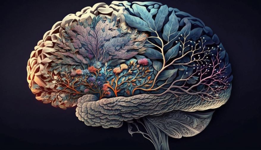
First, during a learning event, a specific subset of neurones becomes highly active. Consequently, these cells express immediate-early genes, which serve as molecular markers of recent activity. Next, researchers can tag these activated neurones using transgenic techniques, allowing them to visualise which cells were engaged. Then, as the event passes and time moves forward, these tagged cells enter a quiet, resting state. However, they remain biochemically altered, making them more likely to fire again in the future.
Preferentially Active Cells Form the Ensemble
Not every neuron takes part in a memory. Sparse groups of cells receive the tag. Their activity during learning predicts selection. Pouget and colleagues deconstructed such an engram. They found multiple ensembles among the tagged cells. Each ensemble tracked distinct features of training. This discovery refines the classic one-trace picture. An engram is really a layered network. Different layers store different details. Context, timing, and emotion each claim a layer. Their collaboration recreates the full experience. This architecture explains memory’s richness. It also explains why recall feels vivid
Synapses Strengthen Where Memory Forms
Engrams take shape through synaptic change. New proteins help connections grow stronger. Repeated firing stabilizes these links. They gain receptors and enlarge with learning. Scientists can measure these physical edits. Spine size increases on tagged dendrites. The changes persist for weeks or more. Sleep deprivation shrinks these spines later. Such findings tie memory to cell biology. Memory is not an abstract ghost. It is a measurable change in wiring. Each strengthened connection adds to the trace.
How Engrams Activate During Recall
Recall begins when a cue reaches the stored trace. The brain matches the cue to past activity. Pattern completion reactivates the original ensemble. Even a partial cue can start the process. Scientists prove this with optogenetics in mice. They label cells that encode a task. Light pulses then trigger the same cells. The animal behaves as if it is recalling.
Light Can Recreate a Stored Experience
Optogenetics gives experimenters a memory switch. Mice learn to fear a safe box. Then researchers flash light at engram cells. The animals freeze as if afraid now. No shock is present during the test. The behavior still appears reliably. This shows engram activation drives recall. Sustained stimulation produced anxiety-like states. Animals showed lasting cellular abnormalities too. The study warns against casual activation. Memory switches carry real emotional weight. Their output shapes mood and behaviour.
Sleep Helps Engrams Mature
Sleep is not passive time for the brain. It replays the day’s important activity. Hippocampal ensembles repeat their waking patterns. These replays occur during slow-wave sleep. The brain then prepares new proteins for storage. Synapses become stronger and more stable. Disturbing sleep interrupts this process. Memory precision drops as a result. The engram remains, but access suffers. Good rest, therefore, guards memory quality. Sleep and memory stay deeply connected.
What Animal Studies Teach Us About Memory
Animal models make engrams testable. Mice offer genetics, imaging, and optogenetics. Researchers condition them to specific contexts. Tagged cells then reveal their storage roles. Threat strength shapes how traces organise. Strong threats recruit more cortical cells. Remote memories depend on these networks. Fruit flies broaden the search further. Their small brains still store usable traces. Each model adds a distinct clue. Together they map how memory works.
Frequently Asked Questions
What exactly is an engram?
First, an engram is a physical trace or biological change in the brain that stores a memory. In simple terms, it is the neural representation of a past experience. Specifically, it consists of a network of neurons that were active during learning and become reactivated during recall.
Who first proposed the engram theory?
Originally, the concept was introduced by German biologist Richard Semon in the early 1900s. However, his ideas were largely ignored at the time because the field leaned more on philosophy than on proof. Later, modern neuroscience confirmed many of his predictions using advanced technology
How do scientists find and tag engram cells in the lab?
First, researchers use transgenic mice engineered to express fluorescent markers in active neurones. For example, they tag cells that turn on immediate-early genes during a learning event. Then, they wait for the event to pass. Later, during recall, the same cells glow again, confirming they form the engram. In addition, tools like calcium imaging and optogenetics allow scientists to watch or control these cells in real time.
Reference:
Otieno, W., Tyukin, I. Y., & Brilliantov, N. (2025). The critical dimension of memory engrams and an optimal number of senses. Scientific Reports, 15(1). https://doi.org/10.1038/s41598-025-11244-y
- Author
- Latest Posts

I’m a web developer and science writer with a Master’s degree in Computer Applications (MCA) from Pune University.
I work in tech, building websites and staying updated with the latest developments. I also love writing about physics, chemistry, technology, robotics, mathematics, and breakthrough innovations. My passion is breaking down complex scientific topics into simple, easy-to-understand stories.
My goal is to help everyone stay informed about the exciting changes happening in science and technology. I believe understanding science shouldn’t be complicated—it should be accessible to all.