The synchronized interplay of the heart's chambers, valves, and electrical conduction system ensures the continuous and efficient circulation of blood.
Cardiac Cycle: Anatomy and Physiology Explained
Every beat of your heart tells a story. In just one day, your heart pumps about 7,600 litres of blood. That is enough to fill nearly 40 bathtubs. This incredible work happens through a precise sequence called the cardiac cycle. The cardiac cycle includes all events from one heartbeat to the next. It involves contraction, relaxation, and pressure changes. Understanding this cycle helps you grasp how your body gets oxygen. It also shows how your body removes waste. This article explains the phases of the cardiac cycle. You will learn about atrial systole, ventricular systole, and ventricular diastole. We will use simple language and clear examples. By the end, you will see your heartbeat as a well-coordinated dance. Let us explore this fascinating process together.
Key Takeaways: Cardiac Cycle
- The cardiac cycle has three main phases: atrial systole, ventricular systole, and ventricular diastole.
- Atrial systole pushes blood from atria to ventricles.
- Ventricular systole pumps blood out of the heart.
- Ventricular diastole allows the heart to refill and rest.
- Pressure changes control valve opening and closing.
- Blood flow follows a one-way path through valves.
- The cycle repeats about 72 times per minute at rest.
- Understanding this cycle helps explain heart health and disease.
What is the Cardiac Cycle? A Simple Overview
The cardiac cycle describes the sequence of events in one heartbeat. First of all, it starts with the contraction of heart muscle. This contraction is called systole. After that, it ends with relaxation, which is called diastole. Above all, the cycle involves pressure changes inside heart chambers. These changes move blood through the heart and into blood vessels. For example, valves open and close based on pressure differences. In addition, the cycle has two major parts: systole and diastole. Each part includes specific events in different chambers. To put it another way, the right side and left side work together. At the same time, they contract at the same instant. The right side pumps blood to lungs. In contrast, the left side pumps blood to the body. As a result, both sides follow the same cycle.
Phase 1: Atrial Systole – The Priming Pump
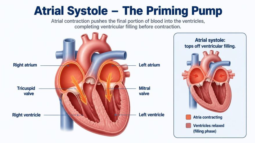
First of all, atrial systole is the first phase of the cardiac cycle. It lasts about 0.1 seconds. During this phase, the atria contract. As a result, this contraction pushes blood into the ventricles. Prior to atrial systole, blood flows passively from veins into atria. Then again, the atria squeeze, adding about 20% more blood to ventricles. Above all, this extra volume is important for heart function. To illustrate, the right atrium receives deoxygenated blood from the body. At the same time, the left atrium receives oxygenated blood from the lungs. After that, both atria contract together. For example, the tricuspid valve opens on the right side. In like manner, the mitral valve opens on the left side. These valves connect atria to ventricles. As can be seen, blood flows down a pressure gradient. At this point, atrial pressure is higher than ventricular pressure.
Phase 2: Ventricular Systole – The Power Phase
Cardiac Cycle: Isovolumetric Contraction
Ventricular systole begins after atrial systole ends. It lasts about 0.3 seconds. The first part is isovolumetric contraction. Ventricles start to contract. Pressure inside ventricles rises rapidly. This pressure exceeds atrial pressure. So the AV valves snap shut. You hear this as the first heart sound, “lub.” All valves are now closed. Volume stays constant inside ventricles. Muscle tension increases. Pressure builds up quickly. No blood leaves yet. This phase is very short. It lasts only about 0.05 seconds. The ventricles prepare to eject blood.
Cardiac Cycle: Ventricular Ejection
Now comes the ejection phase. Ventricular pressure exceeds arterial pressure. The semilunar valves open. The aortic valve opens to the aorta. The pulmonary valve opens to the pulmonary artery. Blood rushes out of ventricles. The left ventricle pumps to the body. The right ventricle pumps to the lungs. Most blood ejects during the first third of ejection. This is called rapid ejection. The remaining blood ejects slowly. This is called reduced ejection. Pressure in ventricles peaks at about 120 mmHg on the left side. On the right side, pressure is lower. It reaches about 25 mmHg. After ejection, ventricular pressure falls. The semilunar valves close. You hear this as the second heart sound, “dub”. This marks the end of systole.
Phase 3: Ventricular Diastole – The Recovery Phase
Cardiac Cycle: Isovolumetric Relaxation
First of all, ventricular diastole is the relaxation phase. It lasts about 0.4 seconds. To begin with, the first part is isovolumetric relaxation. During this time, ventricles relax. As a result, pressure drops sharply. At this instant, all valves are still closed. Consequently, volume remains constant. To put it differently, this phase lasts about 0.08 seconds. After that, when ventricular pressure falls below atrial pressure, the AV valves open. At this point, blood now flows from atria into ventricles. Above all, this phase is critical for heart filling. In fact, without proper relaxation, the heart cannot refill effectively.
Cardiac Cycle: Ventricular Filling
To enumerate, ventricular filling occurs in three stages. First of all, rapid filling begins. During this stage, blood rushes in because pressure is low. As has been noted, about 70% of filling happens here. Second, diastasis occurs. In contrast, this is a slow filling period. At this time, blood flows steadily from veins. Third, atrial systole adds the final 20% of volume. As I have said, you already learned this phase. After that, the cycle then repeats. All things considered, ventricular diastole allows the heart muscle to rest. What’s more, it also allows coronary arteries to fill with blood. To be sure, these arteries supply the heart muscle itself. In conclusion, good diastole is essential for heart health.
Pressure-Flow-Valve Relationships: A Simple Table
First of all, understanding pressure changes helps you visualize the cycle. To illustrate, here is a simple pressure-flow-valve table. As can be seen, the table summarizes key events.
| Phase | Chamber Pressure | Valve Status | Blood Flow |
|---|---|---|---|
| Atrial systole | Atrial > Ventricular | AV valves open, SL valves closed | Atria to ventricles |
| Isovolumetric contraction | Ventricular rising | All valves closed | No flow |
| Ventricular ejection | Ventricular > Arterial | AV valves closed, SL valves open | Ventricles to arteries |
| Isovolumetric relaxation | Ventricular falling | All valves closed | No flow |
| Ventricular filling | Ventricular < Atrial | AV valves open, SL valves closed | Atria to ventricles |
In light of this, the table shows how pressure gradients drive blood flow. For example, valves act like one-way doors. Above all, they prevent backflow. To put it another way, the sequence is precise. As a result, any disruption affects heart function. For instance, if an AV valve leaks, blood flows backward. Consequently, this reduces efficiency. All in all, this table makes the cycle easier to understand.
Frequently Asked Questions: Cardiac Cycle
What is the cardiac cycle?
First of all, the cardiac cycle describes the sequence of events in one heartbeat. To begin with, it starts with the contraction of heart muscle. In other words, this contraction is called systole. After that, it ends with relaxation. To be precise, this relaxation is called diastole. Above all, the cycle involves pressure changes inside heart chambers. As a result, these changes move blood through the heart and into blood vessels.
What is systole?
First of all, systole is the contraction phase of the cardiac cycle. In other words, it is the working phase of the heart. To begin with, systole pushes blood out of heart chambers. For example, atrial systole moves blood into ventricles. After that, ventricular systole forces blood into arteries. As a result, blood travels to the lungs and body. Above all, systole requires strong muscle contractions. To put it differently, the heart squeezes tightly during this phase.
What is diastole?
First of all, diastole is the relaxation phase of the cardiac cycle. In other words, it is the resting phase of the heart. To begin with, diastole allows heart chambers to fill with blood. For example, ventricular diastole follows ventricular systole. After that, the heart muscle relaxes completely. As a result, pressure inside chambers drops sharply. Above all, this pressure drop allows blood to flow in. To put it differently, the heart prepares for the next contraction
References
- Dotter, C. T. (1955). Motion in cardiovascular radiography. Circulation, 12(6), 1034–1042. https://doi.org/10.1161/01.cir.12.6.1034
- Occhetta, E., Corbucci, G., Bortnik, M., Pedrigi, C., Said, S. a. M., Droste, H. T., Hofmann, R., & Marino, P. (2010). Do electrical parameters of the cardiac cycle reflect the corresponding mechanical intervals as the heart rate changes? EP Europace, 12(6), 830–834. https://doi.org/10.1093/europace/euq068
Disclaimer: We do not intend this blog post to provide professional, technical, or medical advice. Therefore, please consult a health professional before making any changes to your diet or lifestyle. In fact, we only use AI-generated images for illustration and decoration. Their accuracy, quality, and appropriate can differ. So, users should avoid making decisions or assumptions based only on the text and image