Queen Mary University of London engineers built SensoPal, a handheld probe that measures how stiff soft material feels and maps hidden lumps in silicone models.
Squeeze a ripe mango and a hard one, and your fingers instantly feel the difference. Doctors use the same trick on the body, and they call it palpation. They press gently on skin to feel for lumps, swelling, or unusually firm spots. Now a Queen Mary University of London team has built a handheld tissue stiffness scanner. It puts a number on that feeling. The device, named SensoPal, turns each press into a stiffness reading. On top of that, a simple camera tracks the probe and draws a map. Saitarun Nadipineni led the work with Mohamed Adhnan Thaha, Kaspar Althoefer, and Thilina Dulantha Lalitharatne. Thaha also works with the Royal London Hospital, part of Barts Health NHS Trust. The paper appeared in Communications Engineering on November 5, 2026, with the DOI 10.1038/s44172-026-00814-1.
Here is the exciting part, stated plainly. In tests with volunteers, the probe measured stiffness with about 87% accuracy. Put simply, its readings scored close to nine out of ten against a lab machine. Its repeatability scored about 86%, so different users got similar numbers. In addition, the color maps flagged deep lumps that people could not feel by hand. Still, one point matters from the very start. All of these tests used silicone models, not real patients. Engineers call such models phantoms, because they stand in for human tissue. The volunteers were engineering students with no medical training. As a result, this work shows a promising prototype rather than a clinic-ready diagnostic tool. Even so, it marks a thrilling step toward fair, measurable touch exams for everyone.
Why Doctors Want a Handheld Tissue Stiffness Scanner
Palpation comes first in many medical checkups, often before any scan. A doctor might press on a belly, a breast, or a neck. Through touch, they judge temperature, tenderness, texture, and stiffness. According to the Communications Engineering paper, this quick check often guides decisions about expensive imaging. Imaging means tools such as CT, MRI, or ultrasound scans. Those scans cost time and money, though. However, touch depends heavily on the examiner’s experience and sensitivity. Two doctors can press the same spot and reach different conclusions. As a result of this, findings stay hard to compare or record. ENTECH recently covered a scoliosis screening suit with sensors with a similar aim. It also replaces guesswork with data. Both projects turn a human impression into a number anyone can check.
The Limits of Judging Lumps by Touch
Some medical machines already measure tissue stiffness inside the body. For example, a method called elastography uses ultrasound or MRI to estimate stiffness. Other optical tools promise even sharper detail. On the other hand, the authors note that such tools are costly and bulky. They rarely suit a small clinic, a home, or a remote village. Earlier handheld devices gave stiffness numbers but could not show a clear visual map. Meanwhile, telemedicine has grown, letting doctors examine patients from far away. Yet a remote doctor cannot reach through a screen and press on a lump. So the team saw a clear gap in everyday health care. They wanted a cheap, portable tool that records stiffness and location together. In this way, a nurse or patient could share data with a distant expert.
What the Volunteer Tests Showed
The team ran four experiments with two groups of sixteen healthy volunteers. Each group contained engineering students from Queen Mary with no prior palpation skills. First, volunteers pressed the probe on six silicone samples of different stiffness. Next, the researchers compared SensoPal’s readings with a commercial materials testing machine. That large lab machine squashes samples at a steady speed and measures the response precisely. The handheld tool reached about 87% accuracy against those reference values. In contrast to that, a perfect copy of the lab machine would score 100%. The team also shared its raw data and code openly for other researchers. Notably, stiffer samples gave more accurate readings than the softest ones. The authors link this to soft silicone acting partly like thick, slow honey. Because of this, very soft samples respond differently to fast and slow presses.
Accuracy Results From the Portable Stiffness Scanner
Repeatability tells us whether different people get the same answer. The portable stiffness scanner scored about 86% on this measure across all sixteen users. Put simply, readings from one volunteer stayed close to readings from another. That matters because real users will not all press the same way. The team also tested whether tilting the probe changed the result. Volunteers held it either flat or tipped at about 30 degrees, a modest angle. Average readings differed only slightly between the two tilt angles. For this reason, the authors judged that the tool works on curved body surfaces too. Additionally, the camera tracked the probe without special lab hardware. A printed square marker on top of the probe guided that tracking. Altogether, these checks suggest the device behaves consistently in ordinary hands, at least on silicone.
Finding Hidden Lumps in Silicone Models
In the next test, volunteers searched a flat silicone block for hidden hard lumps. Each lump sat 3, 6, or 9 millimeters below the surface. Plain silicone measured about 100 kilopascals, a unit of stiffness explained below. In comparison, spots above the 6 and 9 mm lumps read about 200 to 250. That makes them roughly twice as stiff as the plain silicone. On the other hand, the shallow 3 mm lump read about 450. That is nearly four and a half times stiffer. Volunteers found the shallow lump easily, but deeper ones challenged their fingers. By contrast, the stiffness map still highlighted the deeper lumps clearly. A final test used a curved phantom with soft lumps up to 12 mm deep. At 12 mm, volunteers felt the lump only one time in eight. Even so, SensoPal’s readings still told it apart from plain silicone.
Fig. 1: Key Results From The SensoPal Volunteer Tests
Explainer: How the Handheld Tissue Stiffness Scanner Works
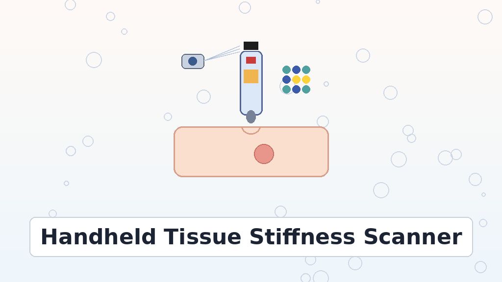
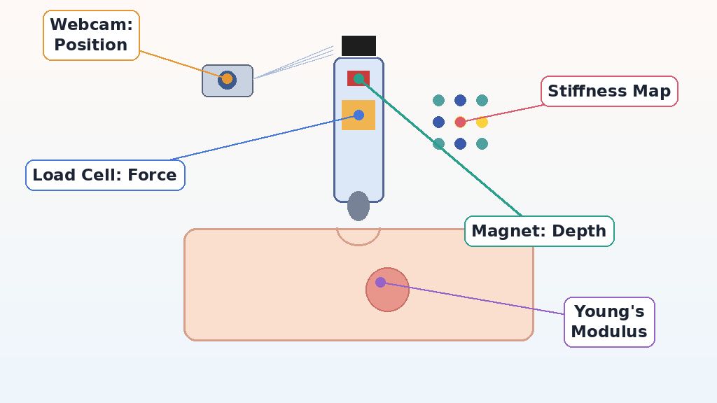
Inside the handheld tissue stiffness scanner sit two simple sensors and a small rounded tip. The tip, about 5 mm in radius, presses into the soft material. Above it, a load cell measures force, much like a tiny kitchen scale. Meanwhile, a small magnet rides up as the tip pushes into the surface. A sensor called a magnetometer feels the magnet’s field grow stronger as it comes closer. In this way, the device works out how far the material has sunk. So every press yields two numbers: how hard you pushed and how deep it went. Then a tiny onboard computer sends both numbers to a laptop. You can explore the basic physics in ENTECH’s guide to elastic behavior in engineering. From there, a classic contact formula converts force and depth into stiffness.
Young’s Modulus in Plain Words
Scientists describe stiffness with a quantity called Young’s modulus. It compares how hard you push a material with how much it squashes. A high Young’s modulus means a stiff material, like a rubber eraser. In contrast, a low value means a squishy material, like jelly. Students studying physics in grade 12 meet this idea with wires and stretching. The unit is the pascal, which equals one newton of force on one square meter. One kilopascal equals a thousand pascals, a handy size for soft tissue. For example, the paper lists stiff muscle between about 5 and 170 kilopascals. Skin, in turn, ranges from about 60 to 850 kilopascals. Because tumors often feel firmer than healthy tissue, stiffness numbers can hint at trouble. Therefore, measuring Young’s modulus makes a doctor’s touch more objective.
How Tissue Stiffness Mapping Builds a Color Picture
A single stiffness number tells you about one spot only. To see a whole area, tissue stiffness mapping links each reading to a location. Here, a low-cost webcam watches a printed square marker on top of the probe. Software then works out where the tip pressed during every push. Next, the program draws a colored dot at that spot on a map. Bright colors mark stiff points, while darker colors mark softer ones. As a result, a hidden hard lump appears as a cluster of bright dots. At the same time, plain areas stay dark and calm. Doctors could later read the map even without touching the patient. Overall, the tool mixes mechanics, magnetism, and computer vision in one compact package. That blend makes it a lively example of engineering serving medicine.
Fig. 2: How A Handheld Probe Measures Tissue Stiffness
Challenges and Motivation Behind the Quantitative Palpation Tool
The motivation for this quantitative palpation tool comes from a fairness problem. Skilled doctors and advanced scanners remain scarce in many parts of the world. According to the authors, an affordable device could widen access to basic checks. A nurse could press the probe while a distant doctor reviews the data. Likewise, patients might one day self-examine at home and send results online. In short, data could travel where doctors cannot. The Queen Mary research repository lists the work under the university’s engineering school. ENTECH has also featured a portable cervical screening device built for village clinics. Building the probe brought real challenges, though. For instance, soft silicone and living tissue react differently to fast and slow presses. As a result, the team matched the lab machine’s speed to typical pressing speed.
What the Study Cannot Show Yet
Every exciting prototype needs honest limits, and this one has several. First of all, the experiments used silicone phantoms, not human patients in a clinic. Real tissue has layers of skin, fat, and organs that behave in complex ways. Also, the volunteers had no medical training, unlike nurses or doctors. On curved phantoms with soft lumps, the maps looked less sharp than on flat ones. The authors suggest better force sensors and smarter signal processing for future versions. In addition, the contact formula works best for small presses. Deep pushes can stretch tissue beyond that simple range and lower accuracy. For this reason, the team plans tests on real biological tissue samples next. Larger clinical studies with patients must follow before anyone uses SensoPal in routine care.
What Comes Next for Point-of-Care Stiffness Checks
SensoPal shows that a handheld tissue stiffness scanner can turn touch into numbers and maps. Not only did it measure silicone well, but it also spotted lumps that fingers missed. That combination could one day support quick checks in clinics, homes, and telemedicine visits. An earlier Research Square preprint shared the first version of this study in 2025. For students, the project links physics, biology, electronics, and coding in one real device. Careers could grow in medical device design, sensor engineering, and clinical testing. In the same way, data scientists may help turn stiffness maps into clear reports. Still, the next big step belongs to patients and real clinics. Those trials will show whether the maps help real doctors. Until those studies arrive, SensoPal remains an inspiring prototype with a bright future ahead.
References
Nadipineni, S., Thaha, M. A., Althoefer, K., & Lalitharatne, T. D. (2026). A handheld tool for Young’s modulus characterization of soft materials: Towards point-of-care tissue assessment. Communications Engineering. https://doi.org/10.1038/s44172-026-00814-1
Nadipineni, S., Thaha, M. A., Althoefer, K., & Lalitharatne, T. D. (2025). A handheld tool for temporal and spatial Young’s modulus characterization of soft materials: Towards point-of-care soft tissue assessment [Preprint]. Research Square. https://doi.org/10.21203/rs.3.rs-7707244/v1
Nadipineni, S., Thaha, M. A., Althoefer, K., & Lalitharatne, T. D. (2025). A handheld tool for Young’s modulus characterization of soft materials: Towards point-of-care tissue assessment (experiment data and codes) [Data set]. Zenodo. https://doi.org/10.5281/zenodo.17048562
Sailee's enthusiasm carries the story, and the piece still keeps the silicone-model limits in plain view. A short side-by-side of how a doctor's fingers and the probe each judge a lump would help younger readers picture the difference. The section on stiffness maps could also say more about who would read the maps and how. For the complete methods and data, readers are encouraged to turn to the original source paper behind this story.
Editorial note
Editor's note: this explainer simplifies a technical engineering study for a general audience, so some detail from the paper is rounded or left out. Anyone curious about the fine points can look them up independently in the original research paper that this story is based on.
Sailee Pakhare holds a Master's degree in Biotechnology, with a research background spanning microbiology, molecular biology, and disease biology. Her postgraduate dissertation examined the antimicrobial potential of bioactive peptides from lactic acid bacteria, and her research experience includes molecular identification of bacterial isolates (with sequencing data submitted to NCBI) and work as a Research Assistant at SafeWright Laboratory. She also completed a summer internship at IIT Hyderabad studying circadian rhythms and disease biology using Drosophila melanogaster as a model organism.
Her work has been recognized with the Best Poster award at the 3rd International Conference on Innovations in Biotechnology Research for Sustainable Bioresources and Bioeconomy (2025), and she has contributed to a book chapter on the role of AI and machine learning in climate change.
Sailee is also a co-founder of LifeXtension, a community focused on longevity, ageing, and health science awareness in India. An interdisciplinary thinker, she explores questions ranging from disease biology to space and the origins of life, approaching science as a way of asking questions rather than a fixed body of facts. As a science communicator and researcher, she is committed to translating complex research into writing that is clear, accurate, and engaging.