Both gram-negative and gram-positive bacteria can resist antibiotics. The outer membrane of gram-negative bacteria makes antibiotic treatment more complicated. They employ mechanisms like efflux pumps or enzymatic deactivation. Efflux pumps push antibiotics out of the…
Gram Positive and Gram Negative Bacteria: Why Their Structures Differ
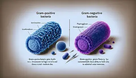
Think of bacteria as tiny warriors surrounded by a protective shield called the cell envelope. First, this structure plays a vital role in bacterial survival because it protects the cell from its surroundings. Furthermore, the cell envelope influences how bacteria respond to antibiotics and other environmental stresses. As a result, differences in the cell envelope can affect whether bacteria survive particular treatments. In 2010, scientists Silhavy, Kahne, and Walker published an important paper that described the bacterial cell envelope and its organisation. Building on this understanding, Mai-Prochnow and colleagues examined another type of stress in 2016: cold plasma. Interestingly, their research showed that Gram-positive and Gram-negative bacteria can respond differently to cold plasma treatment.
Key Takeaways
- Cell Wall Structure: Gram-positive bacteria have a thick layer of peptidoglycan. Gram-negative bacteria have a thin layer plus an outer membrane.
- Stain Result: The Gram stain separates bacteria based on wall thickness. Purple indicates Gram-positive. Pink indicates Gram-negative.
- Sensitivity Factor: Gram-negative bacteria often resist antibiotics better. Their outer membrane acts as a strong barrier.
- Cold Plasma Effect: Gram-positive bacteria are usually more resistant to cold plasma than Gram-negative bacteria. The wall thickness offers protection.
Introduction: Why the Bacterial Cell Envelope Matters
The bacterial cell envelope is much more than a simple outer covering. Instead, it is a complex, multilayered structure that protects the cell from its surroundings and helps maintain its shape. For this reason, understanding the cell envelope is essential for biologists and medical researchers. First, the cell envelope acts as one of the main points of contact between bacteria and external substances, including antibiotics. In addition, many disinfectants target structures within or around this envelope. The 2010 paper by Silhavy, Kahne, and Walker provides an important foundation for understanding these components. In their work, the researchers describe the organisation and functions of the bacterial cell envelope in detail.
The Gram Stain: A Simple Test with Deep Meaning
The bacterial cell envelope is too small to see clearly with a regular light microscope. Therefore, scientists use a laboratory technique called the Gram stain to help distinguish major groups of bacteria. Although scientists developed this method more than a century ago, it remains widely used today. First, scientists apply crystal violet, a purple dye, which initially colours the bacterial cells. Next, they add a mordant, which helps the dye form a more stable complex within the cell envelope. Then, they apply a decolouriser, usually alcohol or an alcohol-based solution. At this stage, the differences between Gram-positive and Gram-negative cell envelopes become important.
The Architecture of Gram-Positive Bacteria
Peptidoglycan Layer
- First, Gram-positive bacteria have a thick peptidoglycan layer, which is one of their main structural features.
- For example, this layer can be compared to a strong, tightly connected mesh that surrounds the cell.
- Structurally, peptidoglycan consists of sugar chains that are cross-linked by short peptide chains.
- Furthermore, this thick layer helps the cell withstand high internal pressure and maintain its shape.
Teichoic Acids
- Next, Gram-positive bacteria contain teichoic acids within and around their thick peptidoglycan layer.
- These polymers contribute to the structure and organisation of the cell envelope.
- Furthermore, teichoic acids contain negatively charged phosphate groups, which influence interactions between the bacterial surface and its surroundings.
- In addition, some teichoic acids are attached to the cytoplasmic membrane. These are known as lipoteichoic acids
The Fortress of Gram-Negative Bacteria
The Outer Membrane
- Now, let us turn to Gram-negative bacteria, which have a more complex cell-envelope architecture.
- First, unlike Gram-positive bacteria, Gram-negative bacteria have a thin peptidoglycan layer, typically only a few nanometres thick.
- However, they also possess an additional outer membrane, which forms a second lipid bilayer around the cell.
- Importantly, this outer membrane acts as a selective barrier between the bacterium and its environment.
The Periplasm
- Next, between the inner (cytoplasmic) membrane and the outer membrane lies the periplasm.
- This region contains the thin peptidoglycan layer and a wide range of proteins and enzymes.
- Furthermore, many periplasmic proteins participate in nutrient transport, processing, protein folding, and other cellular processes.
- In addition, the periplasm contains components involved in electron-transfer processes and helps create a controlled environment for biochemical reactions.
- For example, enzymes in this compartment can begin breaking down certain nutrients before their components are transported into the cell.
Sensitivity to Stress: Cold Plasma and Gram Staining
How Cold Plasma Attacks Cells
The 2016 study by Mai-Prochnow and colleagues tested a new sterilisation method. They used cold atmospheric plasma. This is a gas containing charged particles. It can kill bacteria without high heat. The plasma produces reactive oxygen species (ROS). These are very reactive molecules. They damage the cell membrane. They also break DNA. The study wanted to see if Gram-positive and Gram-negative bacteria reacted the same way. The results were clear. They were not the same. The researchers used Staphylococcus aureus (Gram positive) and Pseudomonas aeruginosa (Gram negative).
Comparison of Sensitivity
The study found that Gram-negative bacteria were more sensitive to cold plasma. This was a surprise to some. Their outer membrane is a strong barrier. But it is also a weak point. The reactive species from the plasma attack the lipids in the outer membrane. This causes the outer membrane to collapse quickly. Once the outer membrane is gone, the cell is vulnerable. The Gram-positive bacteria, with their thick peptidoglycan, survived longer. The mesh was harder to destroy.
Frequently Asked Questions
What is the difference between Gram-positive and Gram-negative bacteria?
The main difference lies in their cell wall structure. Gram positive bacteria have a thick peptidoglycan layer, while Gram negative bacteria have a thin peptidoglycan layer and an outer membrane that acts as a protective shield.
Why do Gram-positive bacteria stain purple in Gram staining?
The thick peptidoglycan layer in Gram-positive bacteria retains the violet dye during the staining process. This gives them their characteristic purple color under a microscope.
Why are Gram-negative bacteria harder to treat with antibiotics?
Their outer membrane blocks many antibiotics from entering the cell. They also have efflux pumps that expel antibiotics and enzymes like β-lactamases that break down drugs, making treatment more challenging
References
- Silhavy, T. J., Kahne, D., & Walker, S. (2010). The bacterial cell envelope. Cold Spring Harbor Perspectives in Biology, 2(5), a000414. https://doi.org/10.1101/cshperspect.a000414
- Mai-Prochnow, A., Clauson, M., Hong, J., & Murphy, A. B. (2016). Gram positive and Gram negative bacteria differ in their sensitivity to cold plasma. Scientific Reports, 6(1). https://doi.org/10.1038/srep38610
- Jordan, S., Hutchings, M. I., & Mascher, T. (2008). Cell envelope stress response in Gram-positive bacteria. FEMS Microbiology Reviews, 32(1), 107–146. https://doi.org/10.1111/j.1574-6976.2007.00091.x
Disclaimer: We do not intend this article/blog post to provide professional, technical, or medical advice. Therefore, please consult a healthcare professional before making any changes to your diet or lifestyle. In fact, we only use AI-generated images for illustration and decoration. Their accuracy, quality, and appropriateness can differ. So, users should avoid making decisions or assumptions based only on the text and images.
- Author
- Latest Posts

Neha Adikane holds a Master’s degree in Environmental Science from Pune University, bringing a unique blend of scientific expertise and creative writing skills to her craft. She specializes in creating engaging, insightful, and impactful content across various niches. With a passion for translating complex ideas into simple, relatable narratives, she excels at crafting blogs, articles, website content, and social media posts that captivate readers and drive engagement. Neha’s background in environmental science adds depth to her writing, allowing her to effectively cover topics related to sustainability, eco-awareness, and scientific innovations.




Your article helped me a lot, is there any more related content? Thanks!