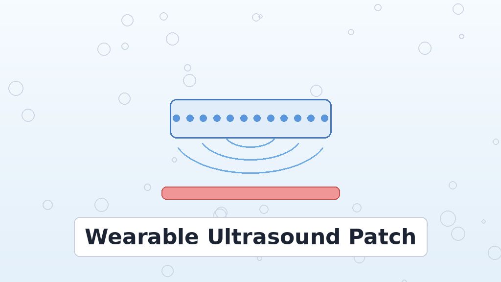
A smartwatch can count your steps and estimate your heart rate. However, it cannot show what happens inside your arteries. Hospital ultrasound can, yet it needs a trained operator and a bulky machine. A wearable ultrasound patch could close that gap. Now, a Chinese team reports a new design. It comes from the Institute of Microelectronics, Chinese Academy of Sciences. Boyuan Wei and Hang Gao led the project. Tsinghua University and the Chinese PLA General Hospital also took part. Their study appears in Microsystems & Nanoengineering under DOI 10.1038/s41378-026-01451-6. In short, the patch runs on only 5 volts. It also images arteries in real time. On top of that, it tracked blood pressure for up to nine hours. As a result, the work hints at hospital-style scans you could wear.
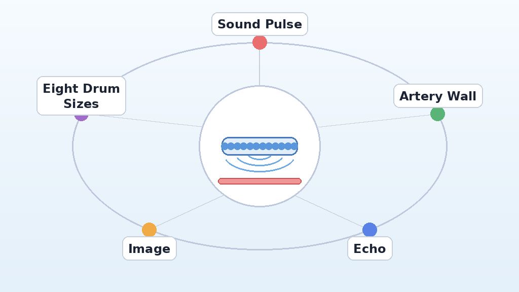
Ultrasound works by sending sound waves into the body and listening for echoes. Most probes rely on chunky ceramic crystals that need high voltage. Because of this, they stay tied to wall power and big electronics. The new device takes a different route. Instead of one large crystal, it uses many tiny vibrating drums on a silicon chip. Engineers call these drums pMUTs, short for piezoelectric micromachined ultrasonic transducers. At the same time, the team mixed eight drum sizes to widen the sound range. That trick gives sharper images without raising the voltage. Still, the authors call the device a proof of concept. Their tests involved only six volunteers. Larger studies must follow. In this article, we explain how the patch works and what comes next.
Why a Wearable Ultrasound Patch Is Hard to Build
Doctors value ultrasound because it is safe, cheap to run, and fast. For example, it shows blood flowing through arteries as it happens. Blood pressure, in particular, links closely to heart health. Our explainer on how the body regulates blood pressure covers the basics. Yet a normal scan lasts only minutes. Pressure, by contrast, changes all day with stress, sleep, and exercise. A cuff reading in a clinic captures one moment only. Therefore, doctors want devices that watch arteries for hours. Wearing a probe sounds simple, but power and size get in the way. Batteries cannot easily supply high voltages for long. Moreover, a thick ceramic probe does not sit comfortably on skin. These limits explain why few ultrasound wearables exist so far. As a rule, more data gives doctors a clearer picture.
High Voltage Keeps Scanners Bulky
Ceramic probes use a material called PZT in bulk form. In practice, they need roughly 10 volts to more than 90 volts to work. Such voltages require bulky driver circuits and careful insulation. For a wearable, that means heavy batteries and extra heat near the skin. Thin-film chips offer an alternative. For this reason, the team chose tiny drums on silicon. Each pMUT drum uses only a thin piezoelectric layer on silicon. Consequently, a small voltage can bend the drum and launch a sound pulse. The returning echo then bends the drum back and creates a signal. Even so, thin drums bring their own trade-off. Each one rings strongly at a single frequency. A narrow frequency range, in turn, limits image detail. So lowering the voltage alone does not solve the whole problem.
Narrow Bandwidth Blurs the Picture
Image sharpness depends on how short each sound pulse is. Short pulses need many frequencies at once, which engineers call wide bandwidth. A single drum, however, acts like a bell with one note. As a result, its pulses ring on and smear fine details together. Earlier pMUT arrays often showed narrow bandwidth for this reason. Some groups added backing layers or extra electronics to fix it. Those fixes, though, often raised cost or voltage again. The new study tackles bandwidth at the design stage instead. In other words, the team shapes the drums so their notes blend. A wider blend of notes means shorter pulses. Shorter pulses, finally, give crisper pictures of artery walls. That idea forms the core of the new framework. Put simply, more notes make a sharper image.
Inside the Chinese Academy of Sciences Design
Tong Jin is the first author of the Microsystems & Nanoengineering study. The team built a linear array of 64 channels in a single row. Each channel holds 24 drum cells grouped into eight sizes. Those cells range from 90 to 125 micrometers across. Because each size rings at its own note, the eight notes overlap. Together, they create one broad band of sound. To plan this, the researchers built an equivalent circuit model of the array. That model also accounts for how neighboring drums affect each other. Thanks to the model, they could predict the final bandwidth before making chips. At just 5 volts, the array reached a fractional bandwidth of 81.7%. That value means the usable frequency range spans a large share of the center frequency.
Merging Frequencies on a Wearable Ultrasound Patch
Picture a choir instead of a solo singer. One voice holds one note, while a choir covers many. Similarly, eight drum sizes give the array many overlapping notes. The team calls this approach frequency merging. In fact, each cell size showed its own resonance peak in air. Once placed against water or tissue, those peaks blurred into one wide response. The fluid damps each drum, which helps the notes merge. Meanwhile, the voltage stayed low, near what a small battery supplies. This balance matters for any wearable design. High bandwidth usually costs either power or size. Here, the team gained bandwidth through geometry alone. So the clever layout, rather than raw voltage, does most of the work. In this way, the patch stays small and light.
Making the Ultrasound Patch on Silicon Wafers
Manufacturing matters as much as clever design. The team built the arrays on 6-inch silicon-on-insulator wafers. Chip factories already handle wafers like these every day. Our guide to how computer chips get made explains the main steps. For example, ion beam etching patterned the top platinum electrode with sharp edges. Thin layers of PZT and platinum then formed each drum. Because many arrays fit on one wafer, the process can scale up. In addition, wafer-level making helps every drum match its neighbors. Uniform drums, in turn, give uniform images. At the same time, standard tools keep the process repeatable. The authors therefore describe their approach as a wafer-scale framework. Still, the study reports lab-scale results only. Mass production would need more testing for yield and long-term reliability.
Fig. 1: Key Results of the Wearable Patch
How a Wearable Ultrasound Patch Sees Inside the Body
Every ultrasound image starts with a tiny sound pulse. The patch sends that pulse into tissue at a few million vibrations per second. When the pulse hits an artery wall, for example, part of it bounces back. The drums then hear this echo and turn it into an electrical signal. Next, a computer measures the delay between pulse and echo. A longer delay means a deeper boundary. By firing many channels in turn, the system builds a full picture. Doctors call this picture a B-mode image. In other words, echo timing becomes a depth map. For instance, the carotid artery sits close to the skin. The group’s earlier low-voltage phased array used similar drums for 3D imaging. This new patch, by contrast, focuses on shallow arteries in the neck and arm.
Echoes, Pixels, and Moving Pictures
A second mode tracks motion over time. In M-mode, the system fires along one line again and again. As a result, it records how an artery wall moves with each heartbeat. The walls swell when blood surges and relax between beats. That wall motion carries clues about pressure inside the vessel. In tissue-like test blocks, the patch resolved details 0.64 millimeters apart in depth. Side-to-side resolution reached 1.27 millimeters. In practice, these numbers describe a fine level of detail. Sound, meanwhile, reached depths of up to 9 centimeters in those test blocks. In people, the team imaged the carotid, brachial, and radial arteries. All three sit within about 4 centimeters of the skin. The paper’s conclusion cites 3 centimeters of in vivo penetration, a small inconsistency worth noting.
Fig. 2: How Echoes Become Artery Images
Testing on Real Arteries and Blood Pressure
Volunteer tests moved the work beyond the lab bench. The patch sat on the neck, upper arm, and wrist of each person. On top of that, it captured clear B-mode and M-mode images at each spot. Wall motion traces then fed a calibration step. For this step, the team used one cuff reading per person. In fact, a cuff still provides the starting point. After that, the patch estimated pressure on its own. Our tips on using a blood pressure cuff correctly show why cuffs stay the reference. Across six volunteers, five men and one woman, results matched a commercial monitor closely. The average deviation reached just 2.2 mmHg against an FDA-approved device. Still, six people form a very small group. Bigger trials will need to confirm it.
Tracking Pressure with Wearable Ultrasound
Long recordings tell a richer story than single readings. In one test, the patch tracked pressure trends for up to nine hours. During that time, the volunteer kept to limited daily postures. In addition, the trends lined up with heart rate and ECG data. That kind of continuous data could reveal spikes a clinic visit might miss. Still, the authors list clear limits. First, the current patch needs better flexibility to hug curved skin. Second, it must resist motion artifacts during walking or exercise. Third, the team wants to shrink the electronics into a CMOS system-on-chip. Finally, larger clinical trials must confirm accuracy in many kinds of people. Until then, the device remains a promising prototype. In short, the path forward looks clear but long.
Conclusion: The Wearable Ultrasound Patch Moves Forward
This study shows that smart geometry can replace brute voltage. By mixing eight drum sizes, the team widened bandwidth to 81.7% at 5 volts. Wafer-scale making, meanwhile, points toward affordable mass production. Volunteer tests then showed sharp artery images and close blood pressure tracking. For now, the wearable ultrasound patch remains a proof of concept with six test subjects. Even so, it tackles two old barriers at once, namely voltage and bandwidth. High blood pressure affects many adults, as CDC heart health guidance notes. Hours of artery data could help doctors spot problems earlier. Later versions may bend with skin and run on tiny chips. In the long run, that could change routine heart care. Someday, sound waves from a sticker could become a daily health tool.
I checked the brief against the published PDF. The device matches the paper: a 1 x 64-channel pMUT array with a frequency-merging design, made on 6-inch SOI wafers, with 81.7% fractional bandwidth at 5 Vp. The numbers are correct: 0.64 mm axial and 1.27 mm lateral resolution, up to 9 cm penetration in phantoms, B-mode and M-mode imaging of carotid, brachial and radial arteries within 4 cm, monitoring up to 9 hours, and 2.2 mmHg deviation against an FDA-approved reference. The six-subject cohort (5 male, 1 female in Fig. 8) and the 10 V to over 90 V range for conventional devices are as stated in the paper. The brief keeps the authors' own limitations on trials, flexibility, integration and power. Verdict: accurate; no corrections needed.
Editorial note
ENTECH runs this story because it brings two big industries together: semiconductor manufacturing and medical devices. Researchers in Beijing made an ultrasound patch on standard silicon wafers that runs on just 5 volts yet images arteries about as well as hospital probes, and it tracked blood pressure for up to 9 hours as volunteers went about daily activities. For teens, it shows how microelectronics, biomedical engineering and acoustics combine in wearable health tech, a field with growing jobs in startups, chip fabs and hospitals. Caveats: only six people were tested, the authors call it a laboratory proof-of-concept, and larger clinical trials, integrated electronics and power measurements are still to come. Takeaway: chip-made, low-voltage ultrasound wearables look feasible, but clinical proof is the next step.
As a seasoned professional, I bring a rich background in strategic planning, institutional effectiveness, and quality assurance to the table. My track record is marked by a proven ability to spearhead the development and implementation of strategic objectives, always with an eye on continuous improvement. I possess deep expertise in managing comprehensive cycles of planning, assessment, analysis, and enhancement of various processes, programs, and services. My adeptness at ensuring adherence to accreditation standards and government regulations is a cornerstone of my professional ethos. My contributions to research have been recognized with an award from the Union Ministry of Energy's Department of Power, underscoring my commitment to excellence and innovation in my field.