Antonie van Leeuwenhoek was a master of optics. Prior to his work, people did not know microorganisms existed. He looked at a drop of pond water.
Antonie van Leeuwenhoek’s Journey Toward Modern Microscopy
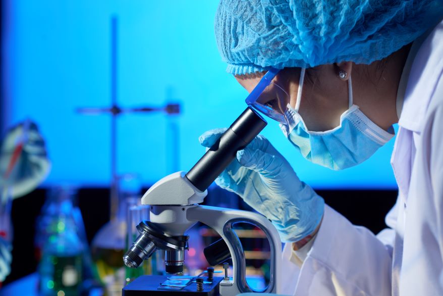
A simple glass bead helped transform biology forever. Indeed, this achievement represents the lasting legacy of Antonie van Leeuwenhoek’s remarkable craftsmanship. During the 1670s, he carefully ground a single lens into a precise, thin curve. As a result, he could observe tiny living organisms that no scientist had previously described. He called these organisms “animalcules”. Unlike many scientists of his time, Leeuwenhoek did not have a university degree. Instead, he developed his skills through careful work with glass and metal. In particular, he sharpened pins, polished glass, and built increasingly powerful microscopes. At first, the Royal Society of London questioned his reports. However, the society arranged for observations to be checked, and subsequently, Robert Hooke and others examined the evidence.
Key Takeaways: Antonie van Leeuwenhoek’s
- Van Leeuwenhoek built over 500 simple microscopes, each with a single, powerful lens.
- His discovery of animalcules proved that life exists at microscopic scales.
- The Delft microscopist’s craft required extreme precision in glass grinding.
- Modern electron and confocal microscopy traces its lineage to Leeuwenhoek’s early methods.
The Delft Lensmaker’s Unique Path
A Draper with a Scientific Eye

Antonie van Leeuwenhoek worked as a cloth merchant in Delft. At first, he used magnifying lenses to inspect thread counts and examine the quality of fabrics. Gradually, this practical skill led him toward microscopy. Although he did not read Latin or Greek, he developed remarkable skill in polishing glass lenses. As a result, he created single-lens microscopes that were small but surprisingly powerful. In particular, each microscope consisted of a silver or brass plate that held a tiny biconvex lens. Meanwhile, the specimen rested on a sharp pin, while screws allowed the user to adjust the focus. With these improvements, Leeuwenhoek’s early microscopes achieved magnifications of up to about 270 times. In comparison, Robert Hooke’s compound microscope generally struggled to provide similarly high useful magnification.
Crafting the Versatile Single Lens
To build his microscopes, first, Leeuwenhoek melted soda-lime glass and pulled it into a thin thread. Next, he reheated the tip to form a tiny glass bead, which became the lens. Afterward, he polished the lens with a special paste. As a result, the process required great patience, precision, and steady hands. Furthermore, Leeuwenhoek placed each lens between two metal plates made of brass or silver. Then, he adjusted the aperture size to control the amount of light entering the microscope. In particular, a smaller opening reduced glare and improved image contrast. Consequently, he could observe microscopic structures more clearly.
The Discovery of Animalcules and Living Cells
First Glimpse of Microbial Life
In 1674, Antonie van Leeuwenhoek collected a water sample from a lake near Delft and placed a drop under his microscope. At first, he noticed green streaks and tiny moving particles. Then, he described the organisms as “animalcules”. Remarkably, he observed that these creatures were about 100 times smaller than a cheese mite. Furthermore, they moved in different ways, including swimming, tumbling, and gliding. In this way, Leeuwenhoek’s early microscopy allowed him to observe microorganisms while they were still alive and active. More specifically, his observations included protozoa and bacteria in their natural environment.
The Narrative Review: A Bridge from Lens to Lab
Bhattacharjee et al. (2024) on the Journey
The review by Bhattacharjee, Singh, and Choudhury (2024) in Applied Microscopy covers van Leeuwenhoek’s life and impact. It traces the evolution from single lens to electron microscope. The authors use original letters and modern references. They show how the Delft microscopist’s craft remained unrivalled for 200 years. The review explains that compound microscopes improved slowly. They suffered from chromatic aberration and blur. In comparison, Leeuwenhoek’s simple lenses stayed sharper and brighter. Only in the 19th century did achromatic lenses catch up. The review highlights that Antonie van Leeuwenhoek’s discoveries spanned 50 years of research. He wrote over 560 letters. He studied over 300 different samples. These included blood, saliva, faeces, and canal water. In detail, the review links his work to modern techniques like phase-contrast and fluorescence microscopy.
The Modern Microscope: Validating van Leeuwenhoek’s Discoveries
Digital Validation with Modern Tools
Today, we can replicate Antonie van Leeuwenhoek’s experiments with digital microscopes and cameras. Scientists have used his original notes to identify the species he saw. For example, the spiral animalcule he drew is likely Spirochaeta. The rod-shaped ones match Bacillus and Escherichia. The single-lens microscope he used could resolve bacteria 2 µm long. That figure is consistent with modern studies. A study by Ramesh et al. (2022) used computer modelling to simulate Leeuwenhoek’s optics. They found that his lenses were nearly perfect spheres. The surface roughness was less than 50 nm. This is extraordinary for a 17th-century craftsman. Above all, it proves that van Leeuwenhoek’s discoveries were accurate and reproducible. The narrative review by Bhattacharjee et al. includes these modern validations. They show that his results hold up under scrutiny.
The Legacy in Biological Sciences
Today, Antonie van Leeuwenhoek’s name appears in almost every microbiology textbook. For this reason, many scientists recognise him as the father of microbiology. More importantly, his observations helped establish the early study of microorganisms and contributed to the development of ideas that later supported germ theory. At the same time, his microscopic observations also helped initiate the study of cells and microscopic life. Furthermore, modern technologies, including confocal and super-resolution microscopy, continue to build on principles of careful lens design and high-quality imaging. In particular, Leeuwenhoek demonstrated how a single, well-made lens could reveal details that scientists could not see with the naked eye. As a result, his approach became an important milestone in the history of microscopy.
Frequently Asked Questions: Antonie van Leeuwenhoek’s
What did Antonie van Leeuwenhoek discover?
He discovered bacteria, protozoa, sperm cells, and blood capillaries. He called them animalcules. His work founded microbiology.
How did Leeuwenhoek’s microscope work?
It used a single, very small glass lens mounted between metal plates. The user held the specimen on a pin and adjusted focus with screws
What is the focus of the narrative review by Bhattacharjee et al. (2024)?
It reviews Leeuwenhoek’s life, his instruments, and his impact on modern microscopy. It connects 17th-century craft to 21st-century science.
Reference:
- Lee, H., Rhyu, I.J.(2026) Antonie van Leeuwenhoek and the dawn of microscopic observation: a narrative review from Delft’s lens to the modern microscope. Appl. Microsc. 56, 3 . https://doi.org/10.1186/s42649-026-00123-z
- Author
- Latest Posts

I completed my Master of Science from the University of Allahabad in 2017 with a strong academic background in life sciences and chemistry. My specialization included Molecular Biology, Microbiology, Genetics, Plant Breeding, Phycology, Paleobotany, and Bioinformatics, along with Organic Chemistry, Inorganic Chemistry, and Physical Chemistry. This multidisciplinary training provided me with a comprehensive understanding of biological systems and analytical scientific approaches.
After completing my postgraduate studies, I gained valuable professional experience in both the education and social development sectors. I worked at A.M. Oxford Public School, where I was actively involved in guiding students toward academic excellence. In this role, I focused on creating an engaging learning environment, encouraging critical thinking, and nurturing students’ curiosity for scientific learning. My experience as an educator strengthened my communication, mentoring, and classroom management skills.
In addition to my teaching experience, I worked with Jeevan Jagriti Foundation, where I contributed to community-based initiatives aimed at improving educational access and awareness among underprivileged sections of society. Through this work, I participated in programs designed to support social development and promote the value of education in marginalized communities.
These professional experiences helped me develop strong interpersonal, leadership, and organizational skills while reinforcing my commitment to education and community service.


