Cellular structural biology connects molecular data with whole-cell structures for deeper understanding. It uses advanced imaging and annotation tools to organize and visualize complex biological data. These methods improve research, support drug design, and enhance…
Cutting Edge Connects Molecular and Cellular Structural Biology
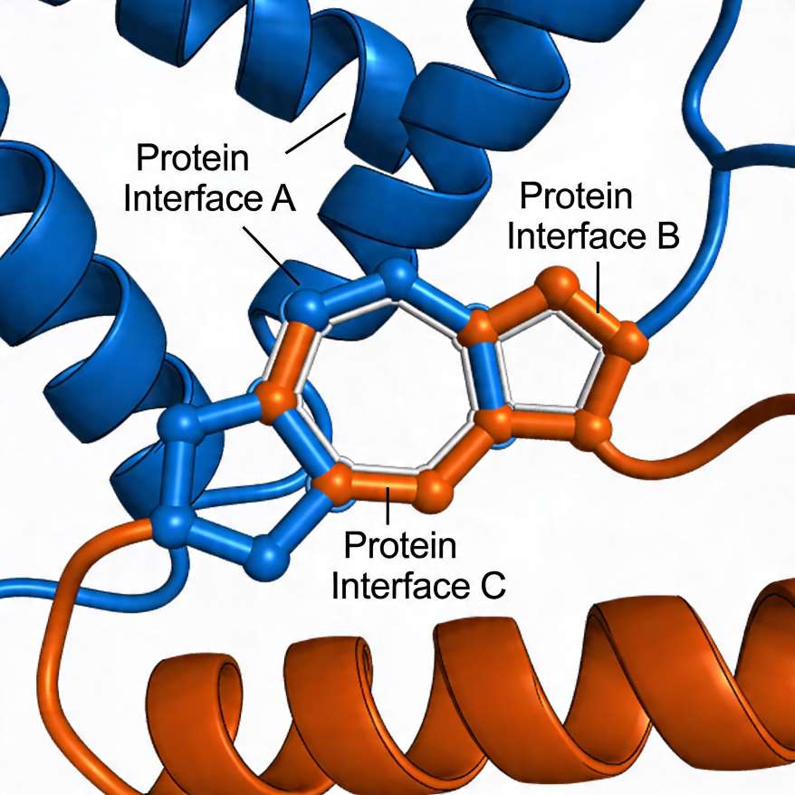
Scientists once saw cells as flat shapes. That view felt simple but not real. As a matter of fact, Cellular structural biology shows cells exist in full three-dimensional space. Today, new tools present clear 3D images of cells. Scientists combine many methods to build these views. At the present time, they connect small molecules with whole cells. To explain, this field links structure with function inside living systems. In this case, proteins move and interact in patterns. These patterns guide health and disease processes. All in all, this work connects school science with real research. It shows how biology and technology grow together.
TL;DR ‘Cellular Structural Biology’
Cellular structural biology connects molecular data with whole-cell structures for deeper understanding. In general, advanced imaging tools create detailed 3D views of cells and their components. To explain, data integration and annotation improve clarity, sharing, and research accuracy. With the result that these methods support better disease study, drug design, and future medical innovation.
Key Takeaways
- Scientists now study cells in full 3D view
- Data from many tools is combined into one system
- This helps explain diseases and cell behavior
- Students can connect school learning to real science
Cellular Structural Biology: Discovery About?
Linking Small Parts to Whole Cells
At first, scientists studied molecules alone. However, cells act as whole systems. To explain, small parts must link with cell context. In this case, scientists mix many tools. These include imaging and data systems. As a result, they build clear 3D models. These models show how parts work together. Another key point, this helps explain disease. It shows what goes wrong in cells. It also helps find better treatments.
Cellular Structural Biology: Link Is Important
In general, data comes from many places. These include the Protein Data Bank and Electron Microscopy Data Bank. However, these sets often stay apart. To put it differently, they need links. With this in mind, new tools connect them. As a result, scientists see a full picture. They understand both shape and function. This makes research faster and clearer.
How 3D Imaging Helps Science in Cellular Structural Biology
Seeing Inside Cells
At the same time, 3D imaging shows more detail. Scientists use methods to split images into small parts. These parts show cell structures. To illustrate, one image becomes many sections. Each part shows a clear feature. These include membranes and proteins. As can be seen, this makes learning easy. Students can understand complex ideas faster.
Main Imaging Tools used in Cellular Structural Biology
- Electron microscopy shows fine cell details; in addition, X-ray methods reveal inner structures.
- To explain, NMR shows how molecules move in space.
- In this case, Cryo-EM studies frozen samples with high clarity.
These tools work together as one system. Each tool adds new detail. Together, they give a full view.
Real Uses in Life
Tools in Cellular structural biology help study viruses in detail. To explain, scientists observe how viruses enter and affect cells. With the result that they design better treatments and understand infection steps clearly. Another key point, drug design improves as researchers study how drugs act on cells. In this case, this knowledge helps create safer and more effective medicines.
Tools That Support Cellular Structural Biology Work
Organizing Biological Data
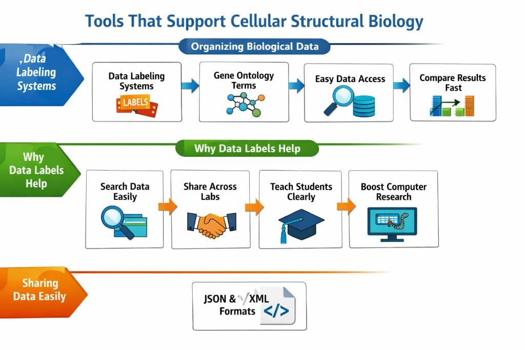
Tools in Cellular structural biology help sort complex data. Scientists use systems that add clear meaning to each dataset. To explain, labels are added to each part using common terms from sources like Gene Ontology Consortium. With the result that data becomes easy to find and use. Another key point, scientists can compare results quickly across studies. In this case, this process supports better teamwork in science.
Why Data Labels Help in Cellular Structural Biology
- Makes data easy to search
- Helps share results across labs
- Supports clear learning for students
- Helps computers study biology
After that, tools make work simple. They guide users step by step. This helps both new and expert users.
Sharing Data Easily in Cellular Structural Biology
Cellular structural biology uses simple formats to store 3D data clearly. To list, formats like JSON and XML keep information neat and organized. With this in mind, these formats support easy handling of complex datasets. At least, this allows smooth data sharing across platforms. In this case, scientists can work together worldwide with better coordination.
Why Cellular Structural Biology Matters for Students
Learning Becomes Clear
At last, Cellular Structural Biology work links school and real science. Students study cells in books. Now they can see real 3D views. To rephrase it, biology becomes easy and visual. It explains cell function and life processes well. As I have said, this builds strong STEM skills. It mixes biology with technology.
Jobs You Can Explore
- Structural biologist
- Bioinformatics expert
- Medical imaging worker
- Data scientist
In short, these jobs use science and data. They solve real problems. Students can follow these paths.
Future Scope
At the present time, research keeps growing. New tools improve each year. Scientists build better systems. Sooner or later, this may change medicine in Cellular Structural Biology aspects.. It may lead to personal treatments. All things considered, this field has great value. It offers many chances for students.
FAQs
What is cellular and molecular structural biology integration?
Cellular structural biology combines molecular data with whole-cell structures. To explain, it links small biological parts with larger systems. With this in mind, scientists understand how cells function in real conditions. In this case, it improves knowledge of biological processes.
Why is segmentation important in 3D imaging?
Segmentation divides images into clear regions. To illustrate, each region represents a specific structure in a cell. With the result that complex data becomes easier to study. Another key point, 3D imaging in Cellular Structural Biology helps visualize biological systems clearly.
What are structural data archives used for?
Archives store biological structure data for research. To list, examples include Protein Data Bank and Electron Microscopy Data Bank. With this in mind, scientists can access shared datasets easily. In short, this supports global scientific collaboration.
Reference
Patwardhan, A., Brandt, R., Butcher, S. J., Collinson, L., Gault, D., Grünewald, K., Hecksel, C., Huiskonen, J. T., Iudin, A., Jones, M. L., Korir, P. K., Koster, A. J., Lagerstedt, I., Lawson, C. L., Mastronarde, D., McCormick, M., Parkinson, H., Rosenthal, P. B., Saalfeld, S., . . . Kleywegt, G. J. (2017). Building bridges between cellular and molecular structural biology. eLife, 6. https://doi.org/10.7554/elife.25835
- Author
- Latest Posts

I completed my Master of Science from the University of Allahabad in 2017 with a strong academic background in life sciences and chemistry. My specialization included Molecular Biology, Microbiology, Genetics, Plant Breeding, Phycology, Paleobotany, and Bioinformatics, along with Organic Chemistry, Inorganic Chemistry, and Physical Chemistry. This multidisciplinary training provided me with a comprehensive understanding of biological systems and analytical scientific approaches.
After completing my postgraduate studies, I gained valuable professional experience in both the education and social development sectors. I worked at A.M. Oxford Public School, where I was actively involved in guiding students toward academic excellence. In this role, I focused on creating an engaging learning environment, encouraging critical thinking, and nurturing students’ curiosity for scientific learning. My experience as an educator strengthened my communication, mentoring, and classroom management skills.
In addition to my teaching experience, I worked with Jeevan Jagriti Foundation, where I contributed to community-based initiatives aimed at improving educational access and awareness among underprivileged sections of society. Through this work, I participated in programs designed to support social development and promote the value of education in marginalized communities.
These professional experiences helped me develop strong interpersonal, leadership, and organizational skills while reinforcing my commitment to education and community service.