Explore the impact of viral lung infection, its symptoms, and how to recognize the need for medical care effectively.
Viral Lung Infection and Its Impact on Lung Health
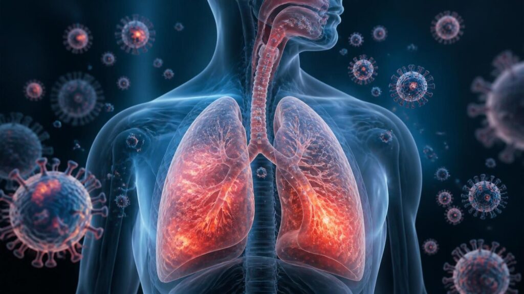
Every year, millions of people worldwide experience these infections. Some recover quickly, while others face severe complications. A viral lung infection occurs when your lungs, which are remarkable organs that work tirelessly every second, are attacked by viruses. They exchange oxygen for carbon dioxide without you even noticing. But when a virus attacks this delicate system, everything changes rapidly. The infection starts in the epithelial cells lining your airways. From there, it triggers a cascade of immune responses. Your body fights back fiercely. This defence mechanism can sometimes cause more damage than the virus itself. Understanding this process helps you recognise symptoms early. It also empowers you to seek proper medical care when needed. In this article, we will explore how viral pathogens invade lung tissue, trigger inflammation, and compromise oxygen exchange. We will discuss the science behind mucus production, alveolar damage, and immune responses. By the end, you will grasp why respiratory viruses pose such serious health risks.
Key Takeaways: Viral Lung Infection
- Viral lung infections begin when pathogens attach to epithelial cells in your airways
- The immune response involves inflammation, mucus production, and cellular battles
- Alveolar damage directly impairs oxygen exchange between air and blood
- Severe infections can lead to pneumonia and acute respiratory distress syndrome
- Early recognition of symptoms improves treatment outcomes significantly
The First Line of Defence: Epithelial Infection and Viral Entry
Epithelial Damage Triggers Alarm Signals
The Battle Within: Immune Response and Inflammation

In the study of viral lung infection, when viruses infect epithelial cells, these cells release danger signals. As a matter of fact, interferons are the first line of antiviral defence. They alert neighbouring cells to prepare for a viral attack. In addition, epithelial cells secrete chemokines that attract immune cells. On the other hand, the illustration of the viral lung infection signalling cascade amplifies the local immune response. Unfortunately, excessive signalling can cause collateral damage to healthy tissue. As a result, the epithelial barrier becomes leaky and compromised. Subsequently, fluid and proteins enter the air spaces. With attention to gas exchange, this accumulation impairs it even further. After all, understanding this mechanism explains why some patients develop severe respiratory distress.
| Virus Type | Cellular Receptor | Primary Target Cells |
|---|---|---|
| Influenza A | Sialic acid | Epithelial cells, ciliated cells |
| SARS-CoV-2 | ACE2 | Alveolar type II cells, endothelial cells |
| RSV | Nucleolin | Ciliated epithelial cells |
| Rhinovirus | ICAM-1 | Nasal epithelial cells |
Role of T Cells and B Cells in Viral Clearance
Adaptive immunity takes several days to develop fully. T cells recognize viral antigens presented by infected cells. CD8+ cytotoxic T cells directly kill virus-infected epithelial cells (Sette & Crotty, 2021). This eliminates the viral replication factories. However, this killing process also destroys functional lung tissue. CD4+ helper T cells coordinate the overall immune response. They help B cells produce specific antibodies against the virus. Neutralizing antibodies prevent viral entry into new cells. Memory B cells and T cells provide long-term protection. Vaccination aims to generate these memory cells without causing disease. The adaptive response ultimately clears the viral lung infection. But the recovery process may take weeks.
The Viscous Problem: Mucus Production and Airway Obstruction
Why Your Lungs Produce Excess Mucus
Mucus is a normal and essential component of lung function. It traps pathogens, dust, and debris. Healthy mucus is thin and easily cleared by cilia. During a viral lung infection, mucus production increases dramatically. Inflammatory cytokines stimulate goblet cells to secrete more mucin. Mucin is the protein that gives mucus its gel-like consistency. The mucus becomes thicker and stickier than normal. This thickened mucus plugs small airways. It creates physical obstruction that makes breathing difficult. Patients cough excessively trying to clear these plugs. But the thick mucus often resists clearance. This leads to persistent coughing and wheezing. In severe cases, mucus plugs can cause atelectasis, or lung collapse.
Mucus Hypersecretion and Airway Remodeling
Chronic mucus hypersecretion causes long-term changes in airway structure. Repeated viral infections can lead to airway remodeling. The smooth muscle surrounding airways thickens. Goblet cell numbers increase permanently (Bonser & Fahy, 2021). This condition resembles asthma-like changes. The airways become hyperresponsive to triggers. Patients experience recurrent wheezing and shortness of breath. Children who suffer severe RSV infections often develop asthma later. The relationship between viral infections and chronic lung disease is complex. Preventing excessive mucus production may reduce long-term damage. Treatment strategies include mucolytics and hydration. Chest physiotherapy helps mobilize thick secretions.
How Mucus Affects Gas Exchange
Mucus accumulation does more than block airways. It directly impairs oxygen exchange at the alveolar level. When mucus covers alveolar surfaces, the diffusion distance increases. Oxygen must travel through a thicker barrier to reach capillaries. This slows down gas exchange significantly. Carbon dioxide elimination is also compromised. Patients develop hypoxemia, or low blood oxygen levels. They may also retain carbon dioxide, leading to respiratory acidosis. Pulse oximetry readings drop below normal levels. Supplemental oxygen may be necessary. In severe viral lung infection, mechanical ventilation might be required. Mucus management becomes a critical component of intensive care.
The Delicate Exchange: Alveolar Damage and Oxygen Impaired
How Viruses Damage Alveolar Cells
At first, viruses infect Type II alveolar cells. These cells have ACE2 receptors on their surface. SARS-CoV-2 uses these receptors to enter cells. Once inside, the virus makes many copies. After that, the cells die and break open. Type I cells are also damaged during infection. As a result, the thin barrier becomes thick. Fluid leaks into the air sacs. To illustrate, this is like water filling up a sponge. The exchange surface becomes smaller. In addition, immune cells rush to the area. They release chemicals that cause more harm. With the result that, healthy tissue gets destroyed.
Alveolar Fluid Accumulation
Fluid buildup is called pulmonary edema. At this point, gas exchange slows down. Oxygen has to travel through extra fluid. This takes more time. So far, we know fluid comes from leaky blood vessels. Viruses damage the cells that line capillaries. Blood plasma escapes into the air spaces. To illustrate, imagine a pipe with holes. Water leaks out everywhere. In like fashion, your alveoli fill with liquid. In short, this prevents oxygen from reaching your blood. Patients feel short of breath. Their oxygen levels drop very low.
Frequently Asked Questions: Viral Lung Infection
What exactly is a viral lung infection?
A viral lung infection, commonly referred to as viral pneumonia or viral bronchitis, occurs when a virus infects the lower respiratory tract, specifically the bronchial tubes and the alveoli (tiny air sacs) in the lungs. First and foremost, this type of infection is distinct from bacterial pneumonia because it is caused by pathogens such as influenza, RSV, or adenovirus rather than bacteria. .
What are the most common symptoms to watch for?
Symptoms typically develop rapidly and can range from mild to severe. For instance, common signs include a persistent dry or productive cough, high fever, chills, body aches, fatigue, and difficulty breathing or chest tightness. However, unlike a bacterial infection, viral lung infections often come with systemic symptoms like headache and muscle pain.
How is a viral lung infection diagnosed?
Diagnosis begins with a physical exam and listening to the lungs with a stethoscope, where doctors may detect crackling or wheezing sounds. A chest X-ray or CT scan is often used to confirm the presence of inflammation or fluid in the lungs, though it cannot always distinguish between viral and bacterial causes.
References
Berg, J., Heinze, J., Niemeyer, D., Hellgren, J., Jaiswal, H., Löwa, A., Hocke, A., Namro, I., Drosten, C., Kurreck, J., & Tolksdorf, B. (2025). Bioprinted Four-Cell-Type Lung Model for Viral Infection Studies Under Air–Liquid Interface Conditions. International Journal of Molecular Sciences, 26(12), 5543. https://doi.org/10.3390/ijms26125543
Stadler, S. V., Von Garnier, C., & Ubags, N. D. (2024). Post-viral lung diseases: the microbiota as a key player. ERJ Open Research, 11(2), 00560–02024. https://doi.org/10.1183/23120541.00560-2024
- Author
- Latest Posts

I completed my Master of Science from the University of Allahabad in 2017 with a strong academic background in life sciences and chemistry. My specialization included Molecular Biology, Microbiology, Genetics, Plant Breeding, Phycology, Paleobotany, and Bioinformatics, along with Organic Chemistry, Inorganic Chemistry, and Physical Chemistry. This multidisciplinary training provided me with a comprehensive understanding of biological systems and analytical scientific approaches.
After completing my postgraduate studies, I gained valuable professional experience in both the education and social development sectors. I worked at A.M. Oxford Public School, where I was actively involved in guiding students toward academic excellence. In this role, I focused on creating an engaging learning environment, encouraging critical thinking, and nurturing students’ curiosity for scientific learning. My experience as an educator strengthened my communication, mentoring, and classroom management skills.
In addition to my teaching experience, I worked with Jeevan Jagriti Foundation, where I contributed to community-based initiatives aimed at improving educational access and awareness among underprivileged sections of society. Through this work, I participated in programs designed to support social development and promote the value of education in marginalized communities.
These professional experiences helped me develop strong interpersonal, leadership, and organizational skills while reinforcing my commitment to education and community service.