Scientists created a new way to keep a special microscope in focus while looking at living things inside the body. This real-time autofocus microscopy method works fast and stays steady even when the animal moves…
Real-time Autofocus Microscopy Enables Robust Intravital Scanning Imaging

Scientists created a new way to keep a special microscope in focus while looking at living things inside the body. This real-time autofocus microscopy method works fast and stays steady even when the animal moves or breathes. It uses a simple trick to check and fix the focus all the time, so the images stay sharp. This lets researchers watch cells and tiny living processes for a long time without stopping to adjust the lens. The result is clear, continuous video of life as it happens inside a living creature.
Key Takeaways
- First, scientists built a new focus tool for microscopes. This tool works very fast. It also stays steady, even when things move. For example, a living animal might breathe or twitch. But the tool still keeps the picture clear because of advances in real-time autofocus microscopy.
- Next, this method is for a special kind of microscope called a light field microscope. It looks deep inside living bodies. Before this, the focus would get blurry over time. Now, the camera can keep recording for a long time thanks to real-time autofocus microscopy.
- Furthermore, the tool checks the focus all by itself. It does not need a person to fix it. This means scientists can watch cells for hours. They do not have to stop to adjust the lens.
- Finally, the result is sharp, clear video. It shows life happening inside a live animal. In short, this new method makes it easy to see tiny things move and change over time.
AI-Based Autofocus Enables Long-Term 3D Live Imaging
Scientists made a new AI tool for microscopes. This tool fixes the focus all by itself. It works very fast. Also, it stays steady even when a live animal moves or breathes. Before this, the picture would get blurry over time. But now, the AI keeps the image sharp. For example, it can look deep inside a living body for many hours. Next, the tool uses a special light field camera. This camera can see cells in 3D. Furthermore, the AI checks the focus again and again. It does not need a person to help. In addition, this means scientists can watch tiny life processes for a long time. Finally, the result is clear, steady video of life inside a live animal. In short, this new method makes long-term 3D live imaging easy and simple by using real-time autofocus microscopy.
High-Speed Autofocus for Live Tissue 3D Microscopy
Scientists created a new way to keep a 3D microscope in focus. This method is very fast. It is called high-speed autofocus, and it is a breakthrough in real-time autofocus microscopy. Second, it works on live tissue inside a living body. For example, an animal might breathe or move. But the focus stays sharp anyway. Before this, the image would get blurry. However, this new tool fixes the focus all by itself. It uses a smart computer program. Furthermore, it checks the focus many times each second. As a result, it never loses the clear picture. Next, this lets scientists watch cells for a long time. They do not need to stop and adjust the lens. In addition, the microscope can see deep inside the tissue. It uses a special light field camera. This camera can make 3D images. Finally, the result is steady, clear video of live tissue moving and changing.
Scanning Light-Field Microscopy Achieves Stable Real-Time Focus
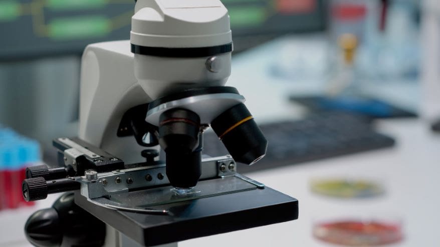
Scientists made a new kind of microscope. It is called scanning light-field microscopy. This tool can keep its focus all by itself. It works in real time. That means it fixes the focus right away, as things happen. For example, a live animal might move or breathe. But the picture stays sharp anyway. Before this, the focus would get blurry over time. However, this new method is stable and steady. It checks the focus again and again. It does not need a person to adjust it. Next, this lets scientists watch cells inside a living body for a long time. They can see 3D images without stopping. In addition, the microscope uses a special light field camera. This camera catches light from many angles. As a result, the images are clear and deep.
Frequently Asked Questions
What is Autofocus for Scanning Light-Field Microscopy?
AFsLF is a new way to keep a microscope in focus. It works on a special kind of microscope called scanning light-field microscopy. This method works in real time. That means it fixes the focus as things happen. First, it watches the focus all the time. It does not need any extra tools or parts. For example, it can keep living tissue sharp for a long time. The tissue might move or breathe. But the picture stays clear anyway. Next, it tracks the focal plane all by itself. It does not need a person to help.
Why is autofocus important in intravital microscopy?
Living tissues move a lot. For example, they move when an animal breathes or when its heart beats. Also, tiny cell activity makes them shift. These small movements can push the sample out of focus. However, AFsLF fixes this problem all by itself. It finds the changes right away. Then it adjusts the focus back. As a result, the image stays sharp. This means researchers can take clear pictures for many days. They do not need to stop and fix the focus. In short, AFsLF keeps the view steady and clear, even with all the natural movement.
What are the main advantages of this autofocus technology?
This method lets scientists take 3D pictures for a long time. It works very fast. Also, it can use many color channels at once. First, it does not harm the living cells very much. That is called low phototoxicity. Next, it works with microscopes that labs already have. You do not need to add new parts or change the hardware. As a result, many labs can start using it right away. In addition, it is easy to set up and use.
Reference
Wang, Y., Wu, J., Wu, J., Li, Y., Lv, W., Yu, F., Yan, J., Lu, Z., Yang, Y., & Dai, Q. (2026). Real-time robust autofocus method enabling sustained intravital scanning light field imaging. Nature Communications, 17. Advance online publication. https://doi.org/10.1038/s41467-026-74976-z
- Author
- Latest Posts

I completed my Master of Science from the University of Allahabad in 2017 with a strong academic background in life sciences and chemistry. My specialization included Molecular Biology, Microbiology, Genetics, Plant Breeding, Phycology, Paleobotany, and Bioinformatics, along with Organic Chemistry, Inorganic Chemistry, and Physical Chemistry. This multidisciplinary training provided me with a comprehensive understanding of biological systems and analytical scientific approaches.
After completing my postgraduate studies, I gained valuable professional experience in both the education and social development sectors. I worked at A.M. Oxford Public School, where I was actively involved in guiding students toward academic excellence. In this role, I focused on creating an engaging learning environment, encouraging critical thinking, and nurturing students’ curiosity for scientific learning. My experience as an educator strengthened my communication, mentoring, and classroom management skills.
In addition to my teaching experience, I worked with Jeevan Jagriti Foundation, where I contributed to community-based initiatives aimed at improving educational access and awareness among underprivileged sections of society. Through this work, I participated in programs designed to support social development and promote the value of education in marginalized communities.
These professional experiences helped me develop strong interpersonal, leadership, and organizational skills while reinforcing my commitment to education and community service.